. 2012 Dec 27;2(6):1657-69.
doi: 10.1016/j.celrep.2012.11.018.

MORC2 signaling integrates phosphorylation-dependent, ATPase-coupled chromatin remodeling during the DNA damage response

Affiliations

MORC2 signaling integrates phosphorylation-dependent, ATPase-coupled chromatin remodeling during the DNA damage response

Da-Qiang Li et al. Cell Rep. .

Abstract

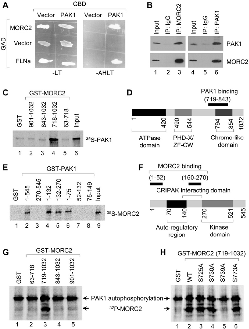
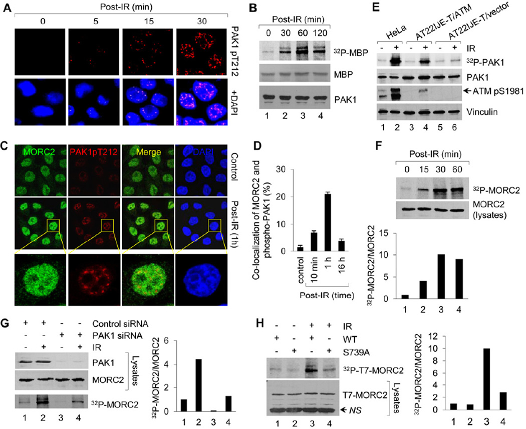
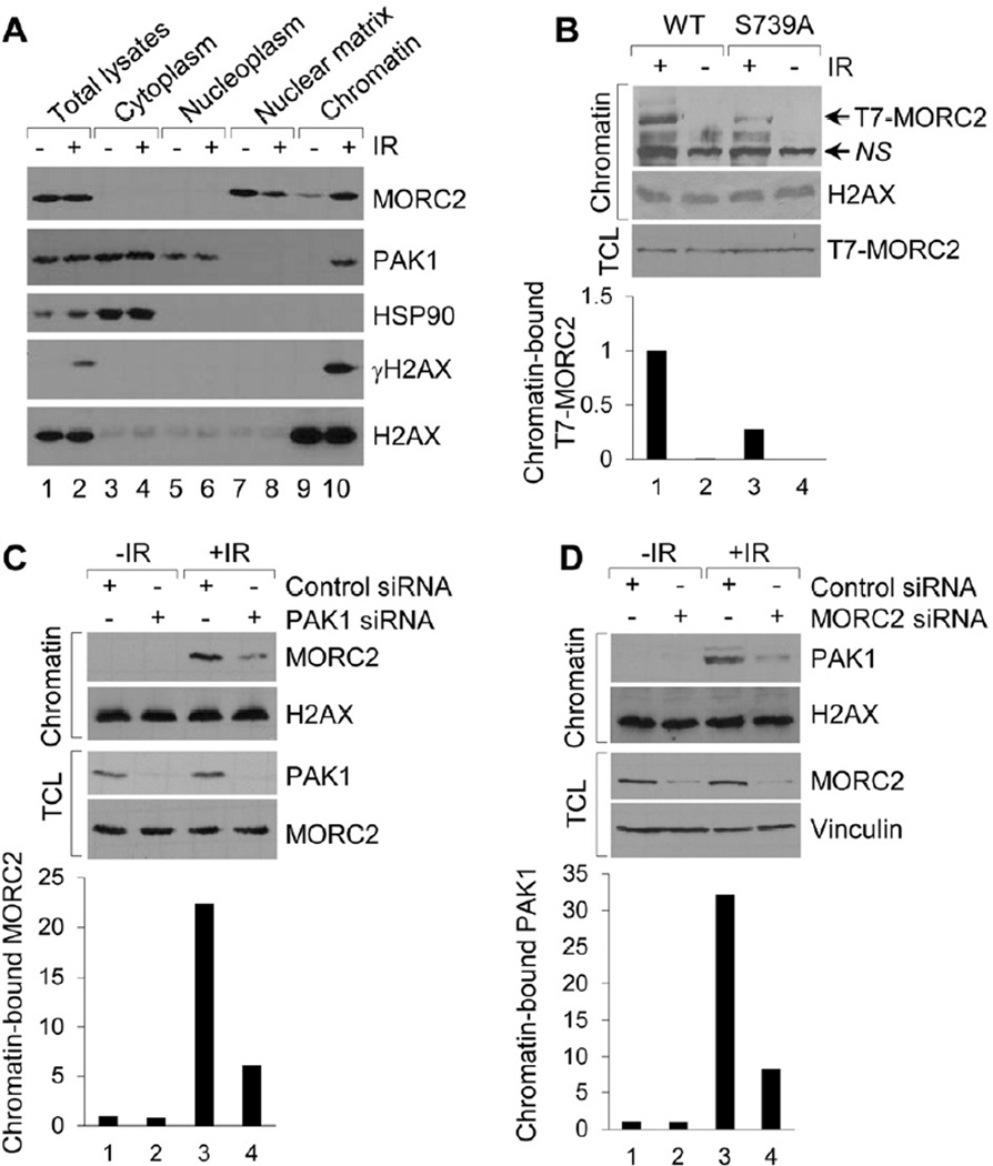
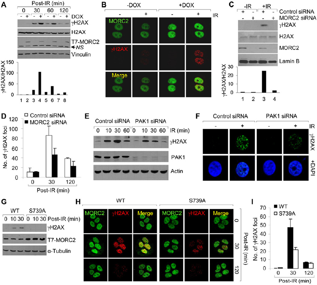
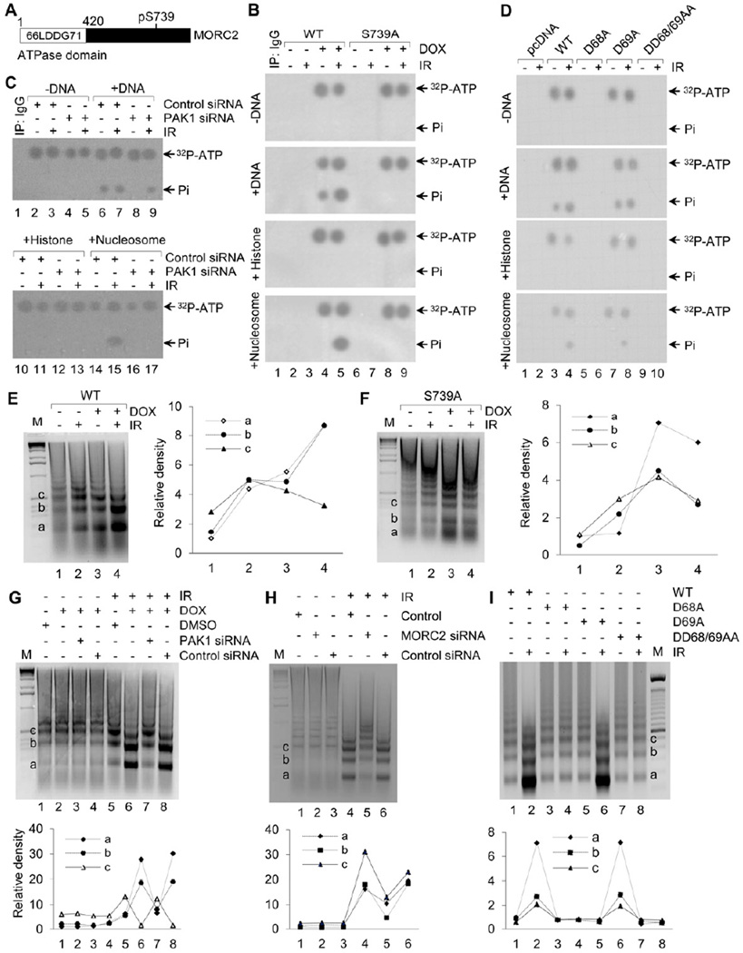
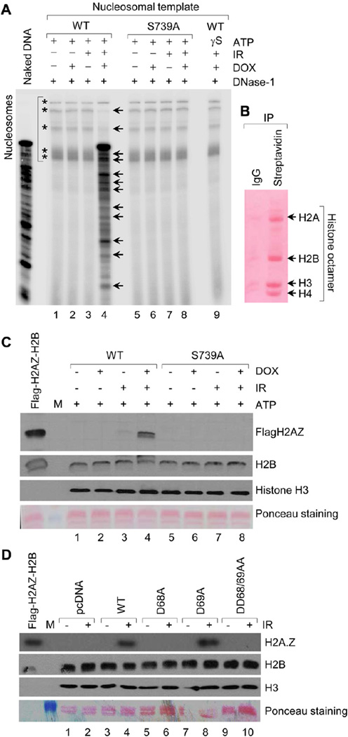
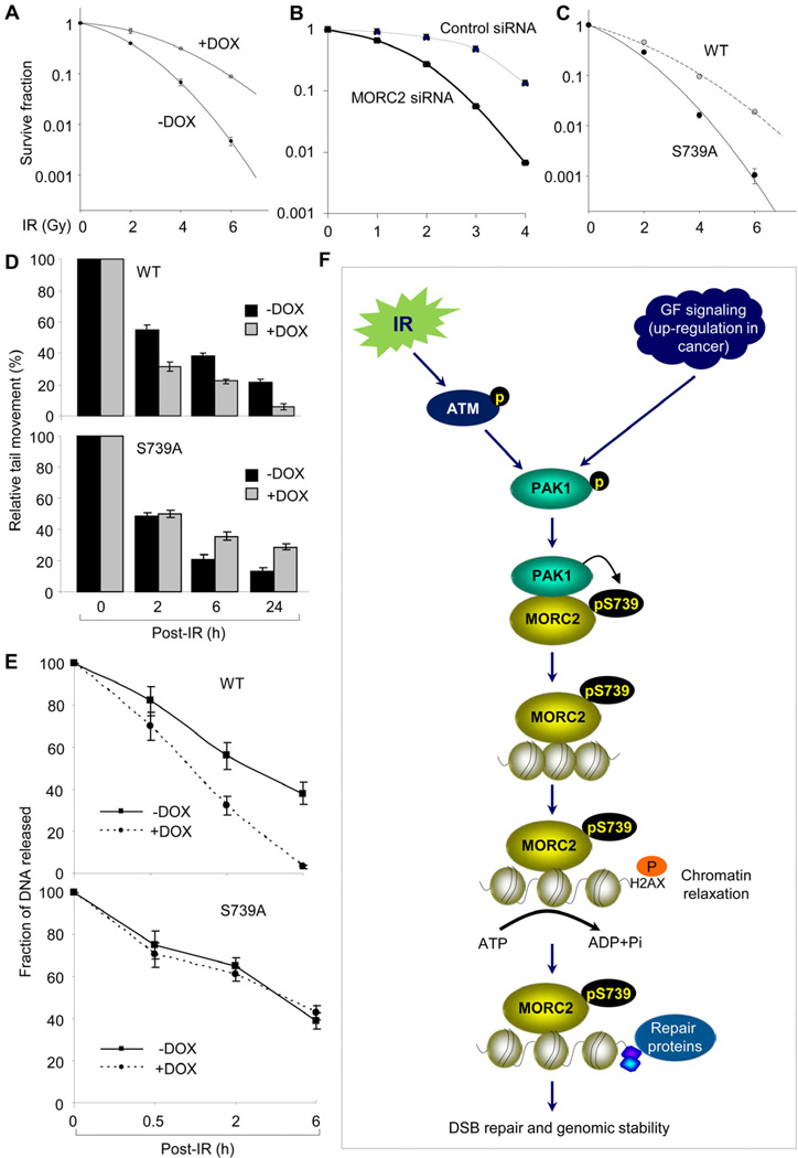
Chromatin dynamics play a central role in maintaining genome integrity, but how this is achieved remains largely unknown. Here, we report that microrchidia CW-type zinc finger 2 (MORC2), an uncharacterized protein with a derived PHD finger domain and a conserved GHKL-type ATPase module, is a physiological substrate of p21-activated kinase 1 (PAK1), an important integrator of extracellular signals and nuclear processes. Following DNA damage, MORC2 is phosphorylated on serine 739 in a PAK1-dependent manner, and phosphorylated MORC2 regulates its DNA-dependent ATPase activity to facilitate chromatin remodeling. Moreover, MORC2 associates with chromatin and promotes gamma-H2AX induction in a PAK1 phosphorylation-dependent manner. Consequently, cells expressing MORC2-S739A mutation displayed a reduction in DNA repair efficiency and were hypersensitive to DNA-damaging agent. These findings suggest that the PAK1-MORC2 axis is critical for orchestrating the interplay between chromatin dynamics and the maintenance of genomic integrity through sequentially integrating multiple essential enzymatic processes.


Figures

Figure 1
Figure 1. MORC2 is A Physiologic Substrate of PAK1 Kinase
(A) Yeast cells were cotransfected with control GAD vector, GAD-MORC2, or GAD-FLNa (filamin-A; positive control) along with GBD vector, or GBD-PAK1. Cotransformants were plated on selection plates lacking leucine and tryptophan (-LT) or adenosine, histidine, leucine, and tryptophan (-AHLT). (B) Lysates from HeLa cells were immunoprecipitated with control IgG, anti-MORC2, or anti-PAK1 antibody, and blotted with the indicated antibodies. (C-D) 35S-labeled, in vitro transcribed-translated PAK1 was incubated with GST-MORC2 deletion constructs and bound proteins were analyzed by SDS-PAGE (C). Schematic representation of the domains of MORC2 for PAK1 binding is shown in D. (E-F) 35S-labeled, in vitro transcribed-translated MORC2 protein was incubated with GST-PAK1 deletion constructs and bound proteins were analyzed by SDS-PAGE (E). Panel F shows the schematic representation of the domains of PAK1 for MORC2 binding. (G) PAK1 phosphorylation of GST-MORC2 deletion constructs by in vitro PAK1 kinase assay. (H) PAK1 phosphorylation of GST-MORC2 (719–1032 amino acids) or its individual mutants.
Figure 2
Figure 2. IR Induces MORC2 Phosphorylation in a PAK1-dependent Manner
(A) HeLa cells were untreated or treated with 10 Gy of IR, harvested at the indicated time points for immunofluorescence staining with the indicated antibodies. Nuclei were visualized by DAPI staining (blue). (B) HeLa cells were untreated or treated with 10 Gy of IR and harvested at the indicating time points. PAK1 immunocomplex was subjected to in vitro kinase assay in the presence of [32P]ATP and MBP as substrates. (C-D) HeLa cells were treated with or without IR and harvested at the indicated time points for immunofluorescence staining of MORC2 and phospho-PAK1 (Thr212). The representative images (C) and quantitation results of co-localization of MORC2 with phospho-PAK1 (D) are shown. (E) Cells were radiolabeled with [32P] orthophosphate overnight. After 1 h of IR treatment, protein extracts were immunoprecipitated with an anti-PAK1antibody, separated by SDS-PAGE, and analyzed autoradiography. Total lysates were immunoblotted with the indicated antibodies for internal controls. (F) HeLa cells were metabolically labeled with [32P] orthophosphoric acid overnight and treated with or without IR. Nuclear extracts were immunoprecipitated with an anti-MORC2 antibody and analyzed by autoradiography. Total lysates were immunoblotted with anti-MORC2 antibody for internal control. (G) HeLa cells were transfected with control siRNA or PAK1 siRNA, metabolically labeled with [32P] orthophosphoric acid overnight. After 1 h of IR treatment, nuclear extracts were subjected to in vivo MORC2 phosphorylation assay. Total lysates were immunoblotted with anti-PAK1 and anti-MORC2 antibodies for internal controls. (H) MCF-7 cells stably expressing wild-type or MORC2-S739A mutant were labeled with [32P]-orthophosphoric acid overnight and treated with or without IR. After 1 h of IR treatment, nuclear extract were immunoprecipitated with anti-T7 agarose beads and subjected to in vivo phosphorylation assay. In F-H, the band densities were quantified using ImageJ software and the results were normalized to the signal for lane 1. NS, non-specific.
Figure 3
Figure 3. MORC2 Associates with Chromatin following DNA Damage in a PAK1 Phosphorylation-dependent Manner
(A) Subcellular fractions were prepared from HeLa cells after 1 h of IR treatment and immunoblotted with the indicated antibodies. (B) Chromatin fractions were isolated from MCF-7 cells stably expressing wild-type or MORC2-S739A mutant after 1 h of IR treatment and then immunoblotted with the indicated antibodies. (C-D) HeLa cells were transfected with control siRNA or specific siRNA targeting PAK1 (C) or MORC2 (D). After 48 h of the second transfection, cells were treated with or without IR. Chromatin fractions were isolated after 1 h of IR treatment and then immunoblotted with the indicated antibodies. In B-D, the band densities were quantified using ImageJ software and the results were normalized to the signal for lane 1. TCL, total cellular lysates; NS, non-specific.
Figure 4
Figure 4. MORC2 Promotes γH2AX Induction in a PAK1 Phosphorylation-dependent Manner
(A-B) MCF-7 cells expressing T7-MORC2 were treated with DMSO or 10 ng/ml of DOX for 12 h and irradiated with or without IR. Cells were harvested at the indicated time points (A) or after 1 h of IR treatment (B), and then subjected to Western blotting (A) or immunofluorescence staining (B) with the indicated antibodies. (C-D) HeLa Cells were transfected with control or MORC2 siRNA. After 48 h of the second transfection, cells were irradiated with or without IR and subjected to immunoblot analysis (C) or immunofluorescence staining (D) with the indicated antibodies. (E-F) HeLa cells were transfected with control or PAK1 siRNA. After 48 h of the second transfection, cells were irradiated with or without IR and then subjected to immunoblot analysis at the indicated time points (E) or immunofluorescence staining after 30 min of IR treatment (F) with the indicated antibodies. (G-I) MCF-7 Cells expressing wild-type or MORC2-S739A mutation were treated with 10 ng/ml DOX for 12 h, irradiated with or without IR, and subjected to immunoblot analysis (G) or immunofluorescence staining (H) with the indicated antibodies. The quantitation results of the immunofluorescence staining of γH2AX foci are shown in panel I.
Figure 5
Figure 5. MORC2 Possesses An Intrinsic ATPase Activity and Facilitates Chromatin Relaxation in Response to DNA Damage
(A) Schematic diagram shows the conserved ATPase domain in the N-terminus of MORC2. (B) MCF-7 cells expressing wild-type MORC2 or S739A mutant were incubated with DMSO or 10 ng/ml of DOX for 12 h. After 1 h of IR treatment, nuclear extracts were immunoprecipitated with T7-tagged agarose beads and subjected to ATPase assays using 100 ng of double-stranded plasmid DNA, histone, or mononucleosomes from HeLa cells. (C) MCF-7 cells expressing wild-type MORC2 were transfected with control or PAK1 siRNA, and treated with or without IR after 48 h of the second transfection. Nuclear extracts were subjected to ATPase assays. (D) HEK293T cells were transfected with the indicated expression vectors. After 48 h of transfection, cells were treated with or without 10 Gy of IR and harvested after 1 h of IR treatment for ATPase assays. (E–F) MCF-7 cells expressing wild-type (E) or S739A mutant (F) were treated with DMSO or 10 ng/ml of DOX for 12 h. After 1 h of IR treatment, nuclear extracts were incubated with 5 units of MNase for 10 min, and DNA was visualized by ethidium bromide staining. (G-H) HeLa cells were transfected with control or specific siRNA targeting PAK1 (G) or MORC2 (H). After 48 h of the second transfection, cells were treated with or without IR and nuclear extracts were subjected to MNase assays. (I) HEK 293T cells were transfected with the indicated expression vectors. After 48 h of transfection, nuclei were prepared after 1 h of IR treatment and subjected to MNase assay. In E-I, the band densities were quantified using ImageJ software and the results were normalized to the signal for a1.
Figure 6
Figure 6. MORC2 Exhibits a Histone-exchange Activity and Promotes Signaling-dependent Nucleosome Mobilization
(A) Nucleosome remodeling assay was carried out using 30 µl of T7-MORC2 or its mutant (S739A) using salt gradient exchange. Total 100 ng of end labeled nucleosomal template was incubated with T7 immunoprecipitates and further digested with DNase1 (0.5U) for 1 min at room temperature. Reaction mix was precipitated, electrophoresed on 10% urea-TBE gel, and exposed to phosphor screen. (B) Schematic for the preparation of native biotinylated chromatin on streptavidin-magnetic beads. (C) Histone exchange assay was carried out using 30 µl of T7-MORC2 with 100 ng of Flag-H2A.Z-H2B dimer and 50 ng of streptavidin bound native chromatin in total of 50 µl exchange reaction at 37 °C for 2 h. Beads were washed with 400 mM KCl and bound proteins were eluted using SDS-Laemmli buffer and analyzed on 16% SDS-PAGE using indicated antibodies. Presence of Flag-H2AZ in elutes demonstrates the exchange of H2AZ for H2A due to chromatin remodeling. (D) HEK293 cells were transfected with the indicated expression vectors. After 48 h of transfection, cells were harvested after 1 h of IR treatment for histone exchange assay as describe above.
Figure 7
Figure 7. MORC2 Promotes DSB Repair and Enhances Radioresistance Following DNA Damage
(A) MCF-7 cells expressing wild-type MORC2 were treated with DMSO or 10 ng/ml of DOX for 12 h, and then subjected to clonogenic survival assays. (B) HeLa cells were transfected with control or MORC2 siRNA. After 48 h of the second transfection, cells were treated with or without IR at the indicated doses and then subjected to clonogenic survival assays. (C) MCF-7 cells expressing wild-type MORC2 or its S739A mutant were treated with DMSO or 10 ng/ml of DOX for 12 h and then subjected to clonogenic survival assays. (D-E) MCF-7 cells expressing wild-type MORC2 or its S739A mutant were incubated with DMSO or 10 ng/ml of DOX for 12 h, and treated with or without IR. Cells were harvested at the indicated time points for comet assays (D) or PFGE assays (E). (F) The proposed working model for MORC2 functions in the DDR. Extracellular signals, such as IR and growth factors (GFs), activate the PAK1 kinase, which interacts with and phosphorylates MORC2 on serine 739. Phosphorylated MORC2 associates with chromatin and facilitates an ATPase-dependent chromatin relaxation in response to DNA damage, which, in turn, might regulate DSB signaling.

References

    1. Bao Y, Shen X. Chromatin remodeling in DNA double-strand break repair. Curr Opin Genet Dev. 2007;17:126–131. - PubMed
    1. Ben-Yehoyada M, Ben-Dor I, Shaul Y. c-Abl tyrosine kinase selectively regulates p73 nuclear matrix association. J Biol Chem. 2003;278:34475–34482. - PubMed
    1. Bochar DA, Savard J, Wang W, Lafleur DW, Moore P, Cote J, Shiekhattar R. A family of chromatin remodeling factors related to Williams syndrome transcription factor. Proc Natl Acad Sci U S A. 2000;97:1038–1043. - PMC - PubMed
    1. Celeste A, Petersen S, Romanienko PJ, Fernandez-Capetillo O, Chen HT, Sedelnikova OA, Reina-San- Martin B, Coppola V, Meffre E, Difilippantonio MJ, et al. Genomic instability in mice lacking histone H2AX. Science. 2002;296:922–927. - PMC - PubMed
    1. Chen CC, Carson JJ, Feser J, Tamburini B, Zabaronick S, Linger J, Tyler JK. Acetylated lysine 56 on histone H3 drives chromatin assembly after repair and signals for the completion of repair. Cell. 2008;134:231–243. - PMC - PubMed

Publication types