

Review
doi: 10.5483/bmbrep.2012.45.12.240.
Structure and catalytic mechanism of human protein tyrosine phosphatome
Affiliations
- PMID: 23261054
- PMCID: PMC4133821
- DOI: 10.5483/bmbrep.2012.45.12.240
Item in Clipboard
Review
Structure and catalytic mechanism of human protein tyrosine phosphatome
BMB Rep.
2012 Dec.
Abstract
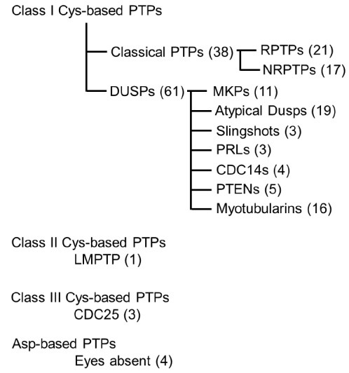
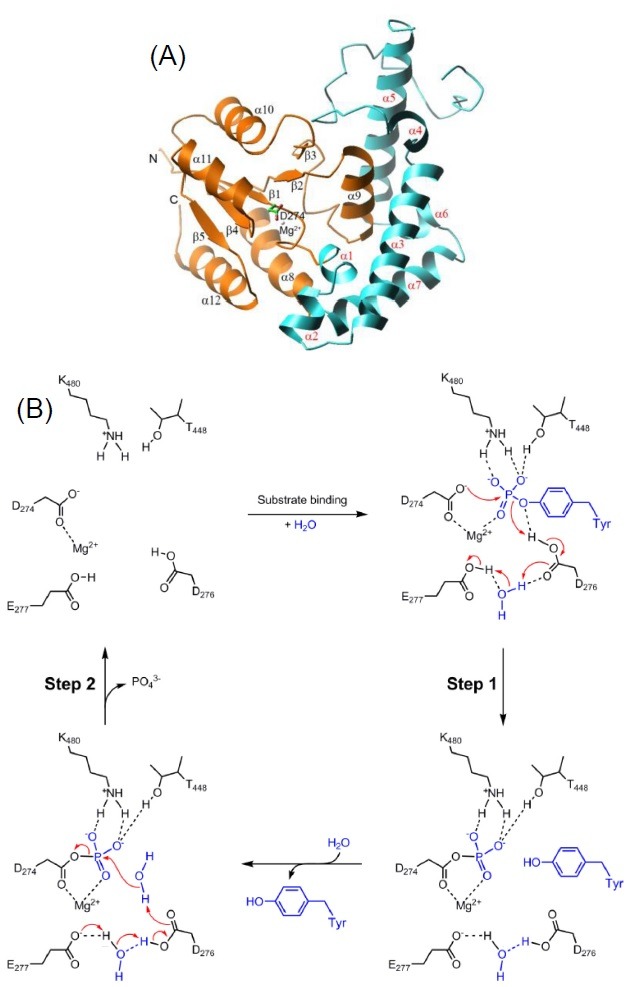
Together with protein tyrosine kinases (PTKs), protein tyrosine phosphatases (PTPs) serve as hallmarks in cellular signal transduction by controlling the reversible phosphorylation of their substrates. The human genome is estimated to encode more than 100 PTPs, which can be divided into eleven sub-groups according to their structural and functional characteristics. All the crystal structures of catalytic domains of sub-groups have been elucidated, enabling us to understand their precise catalytic mechanism and to compare their structures across all sub-groups. In this review, I describe the structure and mechanism of catalytic domains of PTPs in the structural context.
Figures


Similar articles
-
The Extended Family of Protein Tyrosine Phosphatases.Methods Mol Biol. 2016;1447:1-23. doi: 10.1007/978-1-4939-3746-2_1. Methods Mol Biol. 2016. PMID: 27514797 Review.
-
Use of Dominant-Negative/Substrate Trapping PTP Mutations to Search for PTP Interactors/Substrates.Methods Mol Biol. 2016;1447:243-65. doi: 10.1007/978-1-4939-3746-2_14. Methods Mol Biol. 2016. PMID: 27514810
-
An overview of the protein tyrosine phosphatase superfamily.Curr Top Med Chem. 2003;3(7):739-48. doi: 10.2174/1568026033452302. Curr Top Med Chem. 2003. PMID: 12678841 Review.
-
Protein-tyrosine phosphatases: structure, mechanism, and inhibitor discovery.Biopolymers. 1998;47(3):225-41. doi: 10.1002/(SICI)1097-0282(1998)47:3<225::AID-BIP3>3.0.CO;2-O. Biopolymers. 1998. PMID: 9817026 Review.
-
Protein tyrosine phosphatases: structure-function relationships.FEBS J. 2008 Mar;275(5):867-82. doi: 10.1111/j.1742-4658.2008.06251.x. FEBS J. 2008. PMID: 18298793 Review.
Cited by
-
Phosphorylation Dynamics of JNK Signaling: Effects of Dual-Specificity Phosphatases (DUSPs) on the JNK Pathway.Int J Mol Sci. 2019 Dec 6;20(24):6157. doi: 10.3390/ijms20246157. Int J Mol Sci. 2019. PMID: 31817617 Free PMC article. Review.
-
Phosphotyrosine Substrate Sequence Motifs for Dual Specificity Phosphatases.PLoS One. 2015 Aug 24;10(8):e0134984. doi: 10.1371/journal.pone.0134984. eCollection 2015. PLoS One. 2015. PMID: 26302245 Free PMC article.
-
Phosphorylation of Tip60 Tyrosine 327 by Abl Kinase Inhibits HAT Activity through Association with FE65.Open Biochem J. 2013 Aug 22;7:66-72. doi: 10.2174/1874091X20130621002. eCollection 2013. Open Biochem J. 2013. PMID: 24044023 Free PMC article.
-
1H-2,3-dihydroperimidine derivatives: a new class of potent protein tyrosine phosphatase 1B inhibitors.Molecules. 2013 Dec 23;19(1):102-21. doi: 10.3390/molecules19010102. Molecules. 2013. PMID: 24366088 Free PMC article.
-
Allostery in Protein Tyrosine Phosphatases is Enabled by Divergent Dynamics.J Chem Inf Model. 2024 Feb 26;64(4):1331-1346. doi: 10.1021/acs.jcim.3c01615. Epub 2024 Feb 12. J Chem Inf Model. 2024. PMID: 38346324 Free PMC article.
