. 2013 Mar;34(9):2327-39.
doi: 10.1016/j.biomaterials.2012.11.053. Epub 2012 Dec 20.

The effect of the acid-sensitivity of 4-(N)-stearoyl gemcitabine-loaded micelles on drug resistance caused by RRM1 overexpression

Affiliations

The effect of the acid-sensitivity of 4-(N)-stearoyl gemcitabine-loaded micelles on drug resistance caused by RRM1 overexpression

Saijie Zhu et al. Biomaterials. 2013 Mar.

Abstract

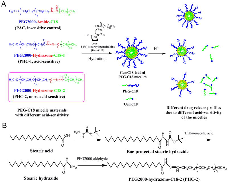
Chemoresistance is a major issue for most gemcitabine-related chemotherapies. The overexpression of ribonucleotide reductase subunit M1 (RRM1) plays a key role in gemcitabine resistance. In this study, we synthesized a new highly acid-sensitive amphiphilic micelle material by conjugating hydrophilic polyethylene glycol with a hydrophobic stearic acid derivative (C18) using a hydrazone bond, which was named as PHC-2. A lipophilic prodrug of gemcitabine, 4-(N)-stearoyl gemcitabine (GemC18), was loaded into micelles prepared with PHC-2, a previously synthesized less acid-sensitive PHC-1, and their acid-insensitive counterpart, PAC. GemC18 loaded in acid-sensitive micelles can overcome gemcitabine resistance, and GemC18 in the highly acid-sensitive PHC-2 micelles was more cytotoxic than in the less acid-sensitive PHC-1 micelles. Mechanistic studies revealed that upon cellular uptake and lysosomal delivery, GemC18 in the acid-sensitive micelles was released and hydrolyzed more efficiently. Furthermore, GemC18 loaded in the highly acid-sensitive PHC-2 micelles inhibited the expression of RRM1 and increased the level of gemcitabine triphosphate (dFdCTP) in gemcitabine resistant tumor cells. The strategy of delivering lipophilized nucleoside analogs using highly acid-sensitive micelles may represent a new platform technology to increase the antitumor activity of nucleoside analogs and to overcome tumor cell resistance to them.


Figures

Fig. 1
Fig. 1
Characterization of PHC-2. (A) 1H NMR spectrum of PHC-2 in CDCl3. (B) 1H NMR spectra of PHC-2 in CDCl3 after incubation up to 24 h at pH 7.4, 6.8 or 5.5, 37°C. a, CHO from PEG-aldehyde, 9.79 ppm; b, CONHN=CH from PHC-2, 8.56 ppm. (C) Time- and pH-dependent degradation profiles of PHC-2 based on 1H NMR spectra in B. (D) The plot of I1/I3 values versus the concentrations of PHC-2 in water (n = 3).
Fig. 2
Fig. 2
In vitro release of GemC18 from PHC-2 micelles in PBS of different pH values (5.5, 6.8, or 7.4) at 37°C (n = 3).
Fig. 3
Fig. 3
Cellular uptake and apoptosis studies. (A) The percentage of GemC18 internalized by TC-1-GR cells after incubation with GemC18, in a solution or in different micelles, for 2 h or 6 h (n = 3). a, P < 0.05, PHC-2 vs. PAC or PHC-1 for 6 h. (B) Representative cytograms of cell apoptosis analysis of TC-1-GR cellsafter treatment with GemHCl, GemC18, GemC18-loaded PAC, PHC-1 or PHC-2 micelles. Untreated cells were used as control. (C) The percentage of different TC-1-GR cell populations (nuclear debris, early apoptotic, late apoptotic and dead, and viable cells) after the treatment with GemHCl, GemC18, GemC18-loaded PAC, PHC-1 or PHC-2 micelles (n = 3).
Fig. 4
Fig. 4
Mechanisms of the cellular uptake of GemC18 in solution or in various micelles. (A) A comparison of cellular uptake GemC18 in solution or in micelles at 4°C vs. 37°C (n = 3). a, P < 0.01 vs. the control. (B) The effect of various specific endocytosis inhibitors on the cellular uptake of GemC18 in three different PEG-C18 micelles (n = 3). b, P < 0.01 vs. the control.
Fig. 5
Fig. 5
A comparison of the intracellular fate of PEG-C18 micelles with different acid-sensitivity. (A) Colocalization of DiO-loaded PEG-C18 micelles (green) and lysosomes (red). Cell nuclei are in blue. Bar, 20 μm. (B–C) The change of FRET effect in TC-1-GR cells. Cells were pre-incubated with DiI/DiO-loaded PEG-C18 micelles for 2 h, followed by six additional hours of incubation in fresh medium. Bar, 20 μm. Values in (C) are derived from the confocal micrographs in B (n = 7–9). a, P < 0.05 vs. PAC; b, P < 0.01 vs. PAC and PHC-1.
Fig. 6
Fig. 6
The effect of NH4Cl on the pro-apoptotic activity of GemC18 in solution or in micelles against TC-1-GR cells and the intracellular degradation of GemC18. The percentage of apoptotic cells after TC-1-GR cells were incubated in the presence (A) or absence (B) of NH4Cl for 24 h. Cells were pre-incubated for 2 h with GemC18, GemC18-loaded PAC, PHC-1 or PHC-2 micelles. Cells pre-incubated with fresh medium were used as a negative control (n = 3). (C) The effect of NH4Cl on the pro-apoptotic activity of the GemC18 in TC-1-GR cells, relative to the negative untreated control. (D) The percentage of GemC18 remaining in TC-1-GR cells 16 h after they were incubated in the presence or absence of NH4Cl after internalization. Cells were pre-treated with GemC18, GemC18-loaded PAC, PHC-1 or PHC-2 micelles for 2 h to allow the internalization of GemC18 (n = 3). a, P < 0.01, 0 mM vs. 50 mM NH4Cl.
Fig. 7
Fig. 7
The effect of different formulations of GemHCl or GemC18 on RRM1 expression and dNTP and dFdCTP levels in TC-1-GR cells. (A) Immunoblotting analysis of RRM1 expression in TC-1-GR cells treated with GemHCl, GemC18, GemC18-loaded PAC, PHC-1, or PHC-2 micelles. Untreated cells were used as the control (Shown are typical data from more than 3 replicates). (B) Cellular dNTP level after treatment with GemHCl, GemC18, GemC18-loaded PAC, PHC-1, or PHC-2 micelles (n = 3). Untreated cells were used as the control. a, P < 0.05 vs. control; b, P < 0.05 vs. GemHCl; c, P < 0.01 vs. GemHCl; d, P < 0.05 vs. control. (C) Cellular dFdCTP level after treatment with GemHCl, GemC18, GemC18-loaded PAC, PHC-1, or PHC-2 micelles (n = 3). e, P < 0.01 vs. GemHCl and GemC18; f, P < 0.01 vs. others. (D) The ratio of dFdCTP to total dNTPs in TC-1-GR cells after treatment with GemHCl, GemC18, GemC18-loaded PAC, PHC-1, or PHC-2 micelles (n = 3). g, P < 0.01 vs. others; h, P < 0.05 vs. others.
Scheme 1
Scheme 1
Schematic representation of micelle formation and the reaction process. (A) Chemical structures of the three PEG-C18 micelle materials, illustration of the preparation of GemC18-loaded micelles, and the expected acid-sensitive release profiles of GemC18 from the micelles. (B) The scheme of the synthesis of PHC-2 conjugate.

References

    1. Barton-Burke M. Gemcitabine: a pharmacologic and clinical overview. Cancer Nurs. 1999;22:176–83. - PubMed
    1. Mackey JR, Mani RS, Selner M, Mowles D, Young JD, Belt JA, et al. Functional nucleoside transporters are required for gemcitabine influx and manifestation of toxicity in cancer cell lines. Cancer Res. 1998;58:4349–57. - PubMed
    1. Bouffard DY, Laliberte J, Momparler RL. Kinetic studies on 2′,2′-difluorodeoxycytidine (Gemcitabine) with purified human deoxycytidine kinase and cytidine deaminase. Biochem Pharmacol. 1993;45:1857–61. - PubMed
    1. Plunkett W, Huang P, Gandhi V. Preclinical characteristics of gemcitabine. Anticancer Drugs. 1995;6 (Suppl 6):7–13. - PubMed
    1. Huang P, Chubb S, Hertel LW, Grindey GB, Plunkett W. Action of 2′,2′-difluorodeoxycytidine on DNA synthesis. Cancer Res. 1991;51:6110–7. - PubMed

Publication types