. 2013 Jan 10;92(1):88-98.
doi: 10.1016/j.ajhg.2012.11.002. Epub 2012 Dec 20.

Splice-site mutations in the axonemal outer dynein arm docking complex gene CCDC114 cause primary ciliary dyskinesia

Affiliations

Splice-site mutations in the axonemal outer dynein arm docking complex gene CCDC114 cause primary ciliary dyskinesia

Alexandros Onoufriadis et al. Am J Hum Genet. .

Abstract

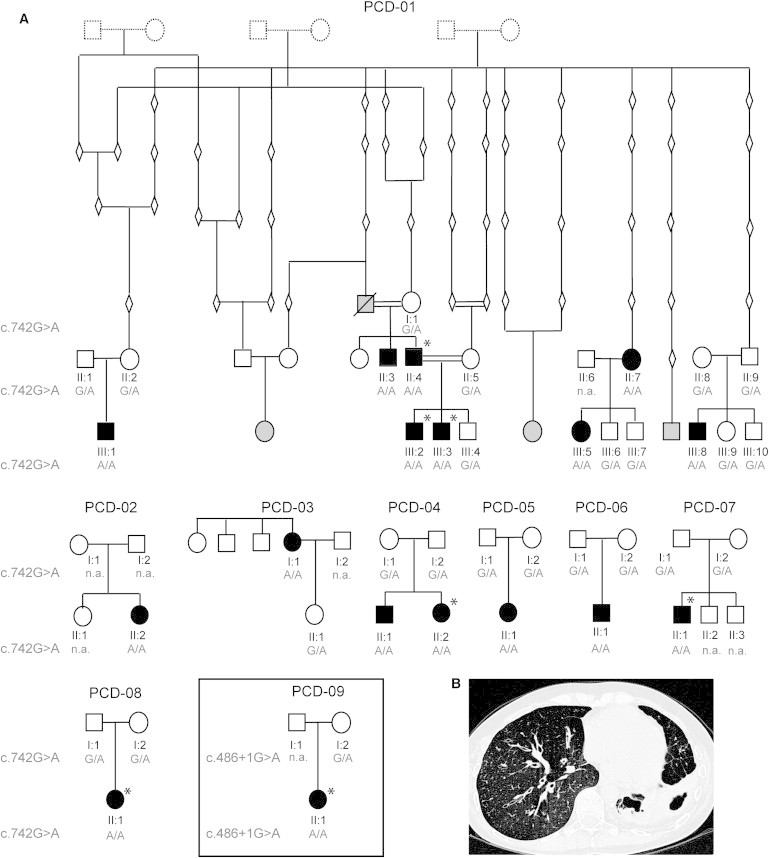
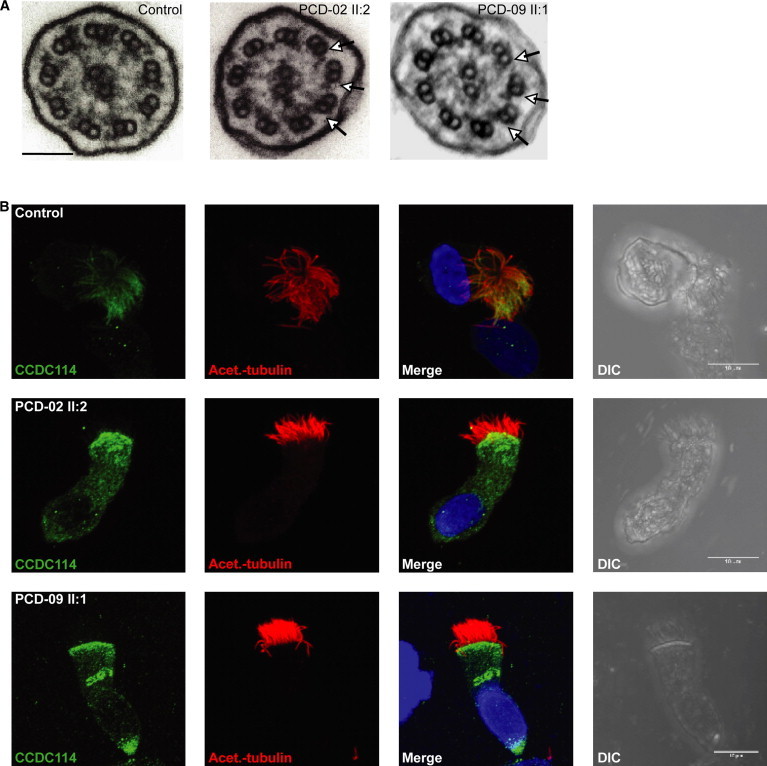
Defects in motile cilia and sperm flagella cause primary ciliary dyskinesia (PCD), characterized by chronic airway disease, infertility, and left-right laterality disturbances, usually as a result of loss of the outer dynein arms (ODAs) that power cilia/flagella beating. Here, we identify loss-of-function mutations in CCDC114 causing PCD with laterality malformations involving complex heart defects. CCDC114 is homologous to DCC2, an ODA microtubule-docking complex component of the biflagellate alga Chlamydomonas. We show that CCDC114 localizes along the entire length of human cilia and that its deficiency causes a complete absence of ciliary ODAs, resulting in immotile cilia. Thus, CCDC114 is an essential ciliary protein required for microtubular attachment of ODAs in the axoneme. Fertility is apparently not greatly affected by CCDC114 deficiency, and qPCR shows that this may explained by low transcript expression in testis compared to ciliated respiratory epithelium. One CCDC114 mutation, c.742G>A, dating back to at least the 1400s, presents an important diagnostic and therapeutic target in the isolated Dutch Volendam population.


Figures

Figure 1
Figure 1
Segregation Analysis of CCDC114 Mutations (A) Pedigree structure of Volendam families PCD-01–PCD-08 showing the segregation of the c.742G>A mutation and of UK family PCD-09 (boxed) showing segregation of the c.486+1G>A mutation. The genealogy of PCD-01 is derived from available church records. Not all ascertained individuals have been shown in the pedigrees, for reasons of space. Filled symbols indicate affected individuals, clear symbols indicate unaffected individuals, gray indicates affected individuals for whom samples could not be obtained, diamonds and dashed symbols indicate confirmed older individuals where samples are unavailable. Asterisks indicate situs abnormalities were reported. (B) High-resolution computed tomography (HRCT) chest scan of an affected Volendam individual showing bronchiectasis of the right and left lower lobes of the lung.
Figure 2
Figure 2
CCDC114 Splice-Site Mutations Causing Primary Ciliary Dyskinesia (A) Effect of the c.742G>A Volendam mutation on splicing. The upper panels show the location of the mutation in genomic DNA sequence chromatograms and the splice-site prediction effect according to Alamut. Alamut uses the four different splice prediction software programs listed on the left. In comparison of the reference sequence from a control individual (top) against the mutant genomic DNA (bottom), the software predicts loss of the splice donor site and presence of a cryptic splice site 79 bp into the intron. The bottom panel shows the sequence of cDNA from a person who is homozygous for the mutation, isolated from ciliary cells and amplified via primers in exons 6 and 8. An intronic insertion of 79 basepairs is present in the c.742G>A individual’s cDNA, located between the mutation substitution site (green arrow) and the presumed intronic cryptic splice site (pink arrow). The sequence shows no indication of use of the regular splice donor site. The inclusion of 79 bases leads to a frameshift and a premature stop codon in exon 8 after addition of 52 novel amino acids, at the in-frame TAA codon indicated by the red box with arrow. (B) Relative expression levels (normalized to ACTB) of CCDC114, CCDC63, and DNAH5 in mRNA from testis and cultured nasal epithelial cells from controls or from Volendam PCD-02 II:2, assessed by qPCR with a Roche Lightcycler as described in Table S5. CCDC114 is expressed at higher levels in cilia-producing cells compared to testis whereas CCDC63 is expressed highly in testis with no detectable expression in cilia-producing cells. In addition, CCDC114 and DNAH5 levels are both reduced in cilia from the Volendam affected individual compared to control. The means ± SEM from triplicate repeat experiments are shown. (C) Location of the Volendam and UK splice-site mutations in the intron-exon structure shown above, and in a model of the CCDC114 protein shown below. Black boxes indicate coding exons, white boxes noncoding exons. The green boxes indicate coiled-coil domains as detected by Paircoil2 run with a minimum window size of 28. Homology was also detected identifying an SMC (structural maintenance of chromosomes protein) domain in CCDC114 indicated by the blue box (SMC_prok_B TIGR02168) and a putative prokaryotic phosphodiesterase domain indicated by the orange box (PRK12704).
Figure 3
Figure 3
CCDC114 Splice-Site Mutations Are Associated with Ciliary Axoneme Defects (A) Transmission electron micrographs of cross-sections of respiratory epithelial cell cilia demonstrates loss of outer dynein arms in both the PCD-02 II:2 and PCD-09 II:1 individuals carrying the c.742G>A and c.486+1G>A splice donor mutations, respectively. All nine peripheral doublets showed loss and reduction of the outer dynein arms (arrows) compared to controls. Scale bar represents 100 nm. (B) Subcellular localization of CCDC114 protein (green) in respiratory epithelial cells via a rabbit polyclonal antibody (Sigma HPA042524). In healthy individuals (top), CCDC114 is localized along the length of the axoneme of the ciliated cells, whereas in both PCD-02 II:2 and PCD-09 II:1, CCDC114 is markedly reduced (middle and bottom). Axoneme-specific anti-acetylated-α-tubulin antibody (Sigma) was used as a control to stain the entire axoneme (red). DNA (blue) was stained with DAPI (Invitrogen). Scale bars represent 10 μm.

Comment in

Similar articles

Cited by

References

    1. Mitchison T.J., Mitchison H.M. Cell biology: how cilia beat. Nature. 2010;463:308–309. - PubMed
    1. Pazour G.J., Agrin N., Walker B.L., Witman G.B. Identification of predicted human outer dynein arm genes: candidates for primary ciliary dyskinesia genes. J. Med. Genet. 2006;43:62–73. - PMC - PubMed
    1. Mitchison H.M., Schmidts M., Loges N.T., Freshour J., Dritsoula A., Hirst R.A., O’Callaghan C., Blau H., Al Dabbagh M., Olbrich H. Mutations in axonemal dynein assembly factor DNAAF3 cause primary ciliary dyskinesia. Nat. Genet. 2012;44:381–389. S1–S2. - PMC - PubMed
    1. Barbato A., Frischer T., Kuehni C.E., Snijders D., Azevedo I., Baktai G., Bartoloni L., Eber E., Escribano A., Haarman E. Primary ciliary dyskinesia: a consensus statement on diagnostic and treatment approaches in children. Eur. Respir. J. 2009;34:1264–1276. - PubMed
    1. Bush A., Chodhari R., Collins N., Copeland F., Hall P., Harcourt J., Hariri M., Hogg C., Lucas J., Mitchison H.M. Primary ciliary dyskinesia: current state of the art. Arch. Dis. Child. 2007;92:1136–1140. - PMC - PubMed

Publication types