. 2013 Mar:241:125-37.
doi: 10.1016/j.expneurol.2012.12.010. Epub 2012 Dec 21.

Anterograde trafficking of neurotrophin-3 in the adult olfactory system in vivo

Affiliations

Anterograde trafficking of neurotrophin-3 in the adult olfactory system in vivo

Huan Liu et al. Exp Neurol. 2013 Mar.

Abstract

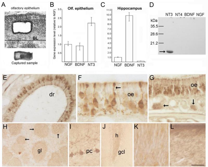
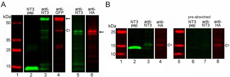
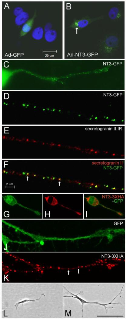
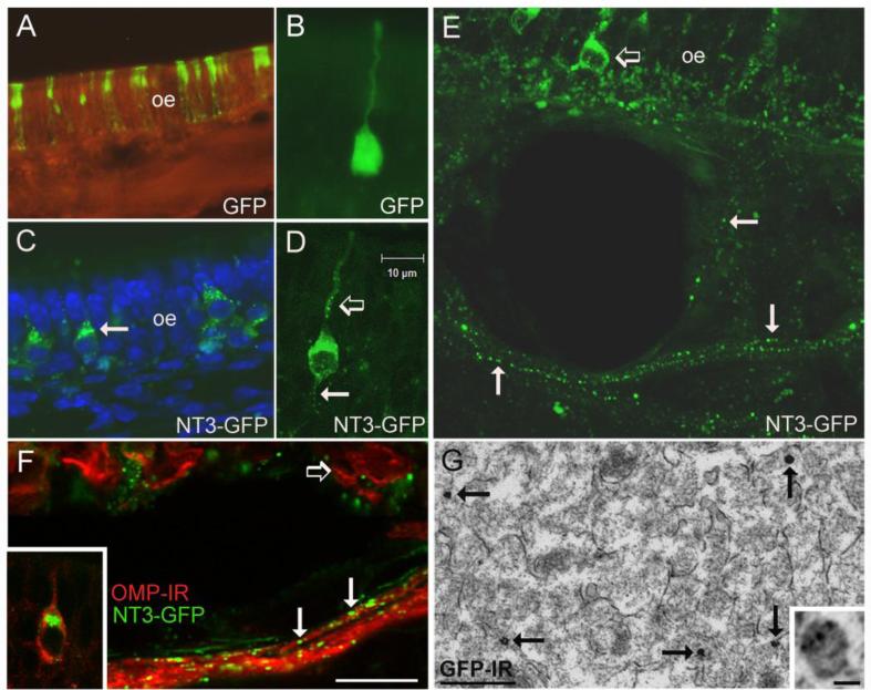
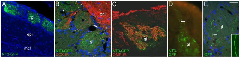
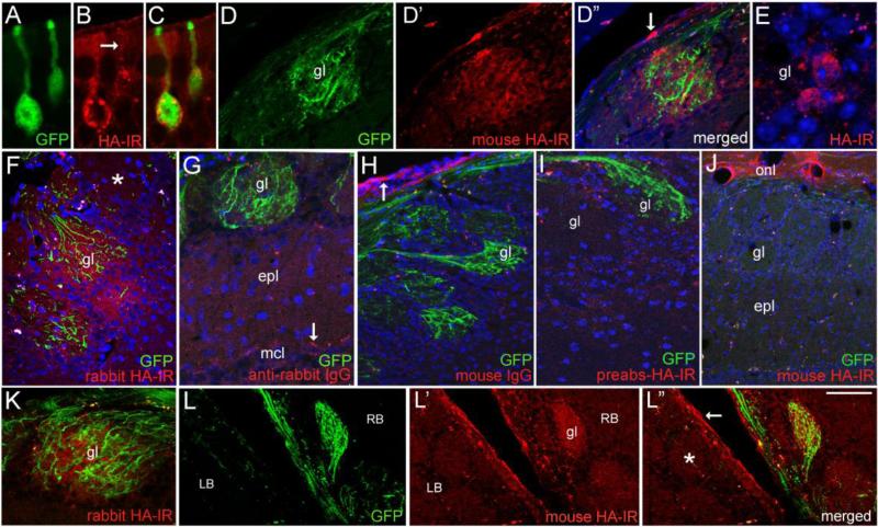
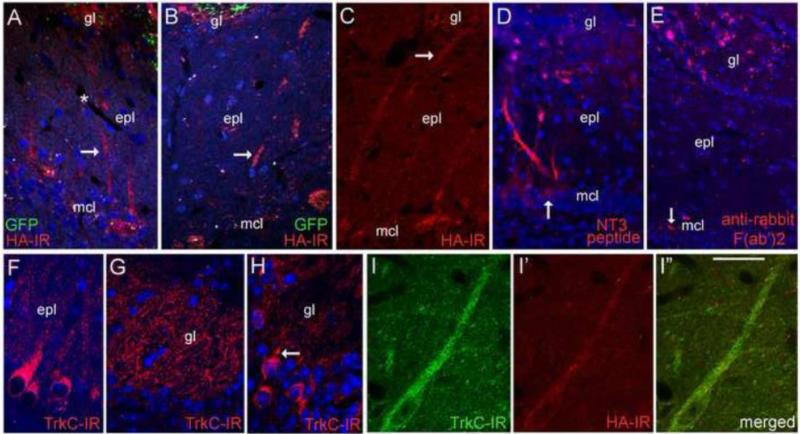
The olfactory system continuously incorporates new neurons into functional circuits throughout life. Axons from olfactory sensory neurons (OSNs) in the nasal cavity synapse on mitral, tufted and periglomerular (PG) cells in the main olfactory bulb, and low levels of turnover within the OSN population results in ingrowth of new axons under normal physiological conditions. Subpopulations of bulb interneurons are continually eliminated by apoptosis, and are replaced by new neurons derived from progenitors in the adult forebrain subventricular zone. Integration of new neurons, including PG cells that are contacted by sensory axons, leads to ongoing reorganization of adult olfactory bulb circuits. The mechanisms regulating this adaptive structural plasticity are not all known, but the process is reminiscent of early nervous system development. Neurotrophic factors have well-established roles in controlling neuronal survival and connectivity during development, leading to speculation that trophic interactions between OSNs and their target bulb neurons may mediate some of these same processes in adults. A number of different trophic factors and their cognate receptors are expressed in the adult olfactory pathway. Neurotrophin-3 (NT3) is among these, as reflected by beta-galactosidase expression in transgenic reporter mice expressing lacZ under the NT3 promoter. Using a combination of approaches, including immunocytochemistry, real-time PCR of laser-captured RNA, and adenovirus-mediated gene transfer of NT3 fusion peptides in vivo, we demonstrate that OSNs express and anterogradely transport NT3 to the olfactory bulb. We additionally observe that in mice treated with adenovirus encoding NT3 tagged with hemagglutinin (HA), a subset of bulb neurons expressing the TrkC neurotrophin receptor are immunoreactive for HA, suggesting their acquisition of the fusion peptide from infected sensory neurons. Our results therefore provide evidence that OSNs may serve as an afferent source of trophic signals for the adult mouse olfactory bulb.


Figures

Figure 1
Figure 1
(A) Photomicrographs of a section through the olfactory epithelium (top) showing the location where cells were isolated by laser microdissection, and the captured cell sample (bottom) as seen on the collection cap. (B-C) Bar graphs showing normalized Q-RT-PCR results obtained from laser-captured OSN samples (B, 45ng total RNA per assay), and dissected hippocampus (C, 20ng total RNA per assay). Mean NGF transcript levels are set to 1.0. Bars indicate mean values calculated for multiple assays, and error bars indicate the range of values obtained across multiple assays for each transcript, rather than statistical error (n=6). (D) Western blot of recombinant neurotrophin peptides (1 μg/lane) demonstrating the specificity of the NT3 antibody. (E-G) Immunostaining for NT3 in the olfactory epithelium (oe). The arrow in F indicates a sensory neuron dendrite, and the arrows in G indicate bundles of more lightly stained sensory axons below the epithelium. (H) In the olfactory bulb, faint staining occurs in the neuropil of glomeruli (gl, and arrows). (I-J) Images showing stained Purkinje cells in cerebellum (I), and more faintly, mossy fibers in the dentate gyrus (J). (K-L) Incubation with pre-absorbed antibody (K) or with pre-immune serum (L) produces non-specific staining in the olfactory epithelium. BL, basal lamina; NL, neuronal layer; SUS, sustentacular cell layer; dr, dorsal recess; gcl, dentate granule cell layer; h, hilus. Bar in K= 40μm in A, 60μm in E, 18μm in F, 41μm in G, 130μm in H, 50μm in I, 100μm in J, and 17 μm in K-L.
Figure 2
Figure 2
In vitro expression of NT3 fusion proteins. (A) Western blots of lysates (15μg protein, lanes 3-6) from infected COS7 cells obtained with Santa Cruz NT3 antibody confirming expression of NT3-GFP (left panel, Santa Cruz anti-GFP), and NT3-3xHA (right panel, Covance anti-HA). Lanes 1 and 2 show molecular weight markers and the signal obtained with 100ng of mature, hrNT3 (control peptide), respectively. Solid arrows indicate the proforms of the tagged trophic factor, and the open arrow indicates a smaller band for mature NT3-GFP. Multiple bands seen for proNT3-3xHA reflect variable glycosylation of the protein. (B) Western blots of COS cell lysates (45μg protein, lanes 3-4) obtained with our rabbit NT3 antibody and Covance mouse anti-HA, showing bands for mature NT3-3xHA (open arrows). Lane 1 shows molecular weight markers and lane 2 shows the signal obtained with 30ng of hrNT3 control peptide. Control lanes (6-8) demonstrate that preabsorption of the NT3 antibody with hrNT3 eliminates detection of hrNT3 (30ng, lane 6) and HA-tagged mature NT3 (lane 7), while the HA epitope is still detected (open arrow, lane 8).
Figure 3
Figure 3
In vitro localization and bioactivity of NT3 fusion proteins. (A-B) Localization of GFP in COS cells infected with Ad-GFP (A) or Ad-NT3-GFP (B). The arrow in B indicates the fusion protein concentrated in the Golgi apparatus. (C) N2a cell infected with Ad-NT3-GFP showing punctate distribution of GFP within a neurite. (D-F) Confocal images illustrating co-localization of NT3-GFP with secretogranin II-IR (arrows in F) in a neurite extending from a cultured N2a cell. (G-K) N2a cells infected with NT3-3xHA-IRES-GFP contain GFP throughout (G), and HA-IR concentrated in the secretory apparatus, including a vesicular-like distribution within neurites (arrows in K). (L-M) Comparison of DRG neuron morphology at 4 days after treatment with conditioned medium from COS cells infected with Ad-GFP (L) or with Ad-NT3-3xHA-IRES-GFP (M). Bar in A= 20 μm for A-B. Bar in F= 2 μm for D-F, J-K. Bar in M= 20 μm in C, 42 μm in G-I, 100 μm in L-M.
Figure 4
Figure 4
In vivo infection of olfactory sensory neurons. (A) Expression of GFP in a portion of the olfactory epithelium (oe) at 5 days after irrigation with the Ad-GFP control virus. The GFP label indicates both sensory neurons and sustentacular cells are infected. (B) An olfactory sensory neuron infected with Ad-GFP showing diffuse fluorescence throughout the cell. (C) Distribution of GFP in sensory neurons expressing NT3-GFP. The arrow indicates fluorescence concentrated in the Golgi apparatus. (D) A sensory neuron expressing NT3-GFP at 5 days after infection. Note the punctate distribution of GFP in the dendrite (open arrow) and in the proximal axon segment (arrow). (E) The vesicular-like distribution of NT3-GFP (arrows) can be followed in sensory axons exiting the epithelium. The open arrow indicates a GFP+ sensory neuron. (F) NT3-GFP+ puncta (arrows) are associated with olfactory sensory axons immunoreactive for OMP (red). The open arrow indicates OMP+ neurons in the epithelium, and the inset illustrates NT3-GFP concentrated in the Golgi apparatus of a mature, OMP+ sensory neuron. (G) Electron micrograph showing the distribution of GFP-immunogold-silver labeling in sensory axons in the olfactory nerve layer of a mouse treated with Ad-NT3-GFP. The arrows indicate dense gold-silver deposits, and the inset shows smaller deposits associated with a presumptive secretory vesicle that is less heavily labeled. Bar F= 58μm in A, 15μm in C, 20μm in E, 10μm in F. Bar in D=10μm in B and D. Bar in G= 500nm, inset=40nm.
Figure 5
Figure 5
Anterograde transport of NT3-GFP to the olfactory bulb. (A) NT3-GFP is distributed in bulb glomeruli (gl) at 5 days after sensory neuron infection. (B) Higher magnification image of glomeruli showing the punctate nature of the NT3-GFP label. The section is immunostained with doublecortin antibody to show the overlying olfactory nerve layer (onl). (C) The distribution of NT3-GFP and axonal OMP-IR overlaps within individual glomeruli. (D) Ad-NT3-GFP infected OSNs project axons containing vesicular-like GFP (arrow) to bulb glomeruli. (E) In contrast, neurons infected with control Ad-GFP project axons in which the GFP signal is distributed uniformly (inset). Epl, external plexiform layer; mcl, mitral cell layer. Bar in F= 35μm in A, 23μm in B-C, 14μm in D, 30μm in E, 7μm in inset.
Figure 6
Figure 6
Expression and distribution of NT3-3xHA in vivo. (A-C) Confocal images comparing the distribution of GFP and mouse HA-IR in sensory neurons 5 days post-infection (d.p.i.) with Ad-NT3-3xHA-IRES-GFP. Vesicular-like distribution of HA is seen in the dendrite (arrow). (D-D”) HA-IR is detected in a glomerulus (gl) containing GFP+ axons. The arrow in D” indicates non-specific labeling of ensheathing glia in the outer bulb nerve layer. (E) HA-IR in cell bodies bordering a glomerulus. HA-negative cells are present as well. (F) Glomerular staining produced by rabbit anti-HA antibody (Cell Signaling) at 7 d.p.i. The asterisk indicates a nearby glomerulus that lacks both GFP+ fibers and HA-IR. (G-I) Staining controls showing lack of HA-IR in sections processed without the rabbit primary antibody (G), incubated in mouse IgG (H), or treated with preabsorbed mouse anti-HA (I). The arrow in G indicates autofluorescent particles in mitral cells, and the arrow in H indicates non-specific labeling of ensheathing glia and processes by mouse IgG. (J) A section from the mouse shown in D, showing a bulb area lacking GFP+ axons. Glomerular HA-IR is also lacking, but non-specific staining of ensheathing glia processes occurs in the olfactory nerve layer (onl). (K) A GFP+/HA+ glomerulus at 7 d.p.i (6.5μm optical section thickness). (L-L”) Low magnification image of a horizontal section showing the medial right (RB) and left bulbs (LB) at 7 d.p.i. A GFP+ glomerulus (gl) in the right bulb is also HA+. GFP and HA-IR are lacking in glomeruli in the left bulb (asterisk). Non-specific staining occurs in the outer nerve layer (arrow in L”). epl, external plexiform layer; mcl, mitral cell layer. Bar in L”= 12μm in A-C and E, 40μm in D-D” and G, 38 μm in F and I, 64 μm in H, 80μm in J and L-L”, and 34 μm in K.
Figure 7
Figure 7
Bulb neuron accumulation. (A-C) Mitral cells deep to GFP+/HA+ glomeruli exhibit HA-IR. Arrows indicate dendritic processes. The asterisk in A indicates a blood vessel in the external plexiform layer (epl). (D). Labeled bulb neurons are seen 3 hrs after glomerular application of AF568-conjugated NT3 peptide. The arrow indicates a labeled mitral cell body. (E) Mitral cell labeling is not observed after glomerular layer injection of AF568-conjugated F(ab')2 fragments, but fluorescent particles are distributed near the injection site. The arrow indicates autofluorescent particles in the mitral cell layer (mcl). (F-H) TrkC-IR localizes to bulb neurons, including mitral cells and their dendrites (F), dendritic processes within glomeruli (gl, G), and scattered cells located in the glomerular layer (H). The arrow in H indicates a glomerular cell with a proximal dendrite directed toward a glomerulus (I-I”) Confocal images showing HA-IR in a TrkC+ tufted cell near a GFP+/HA+ glomerulus at 5 days after intranasal Ad-NT3-3xHA-IRES-GFP treatment. Bar in I” = 46μm in A, 37μm in B, 53μm in C, 56μm in D, 62μm in E, 27μm in F, 21μm in G, and 29μm in H-I”.

Similar articles

Cited by

References

    1. Adachi N, Kohara K, Tsumoto T. Difference in trafficking of brain-derived neurotrophic factor between axons and dendrites of cortical neurons, revealed by live-cell imaging. BMC Neurosci. 2005;6:42. - PMC - PubMed
    1. Alonso M, Viollet C, Gabellec MM, Meas-Yedid V, Olivo-Marin JC, Lledo PM. Olfactory discrimination learning increases the survival of adult-born neurons in the olfactory bulb. J Neurosci. 2006;26:10508–13. - PMC - PubMed
    1. Alvarez-Borda B, Haripal B, Nottebohm F. Timing of brain-derived neurotrophic factor exposure affects life expectancy of new neurons. Proc Natl Acad Sci U S A. 2004;101:3957–61. - PMC - PubMed
    1. Ardiles Y, de la Puente R, Toledo R, Isgor C, Guthrie K. Response of olfactory axons to loss of synaptic targets in the adult mouse. Exp Neurol. 2007;207:275–88. - PMC - PubMed
    1. Baj G, Leone E, Chao MV, Tongiorgi E. Spatial segregation of BDNF transcripts enables BDNF to differentially shape dendritic compartments. Proc. Natl. Acad. Sci. USA. 2011;108:16813–8. - PMC - PubMed

Publication types

MeSH terms