

Abundant pyroglutamate-modified ABri and ADan peptides in extracellular and vascular amyloid deposits in familial British and Danish dementias
- PMID: 23261769
- PMCID: PMC3570712
- DOI: 10.1016/j.neurobiolaging.2012.11.014
Abundant pyroglutamate-modified ABri and ADan peptides in extracellular and vascular amyloid deposits in familial British and Danish dementias
Abstract
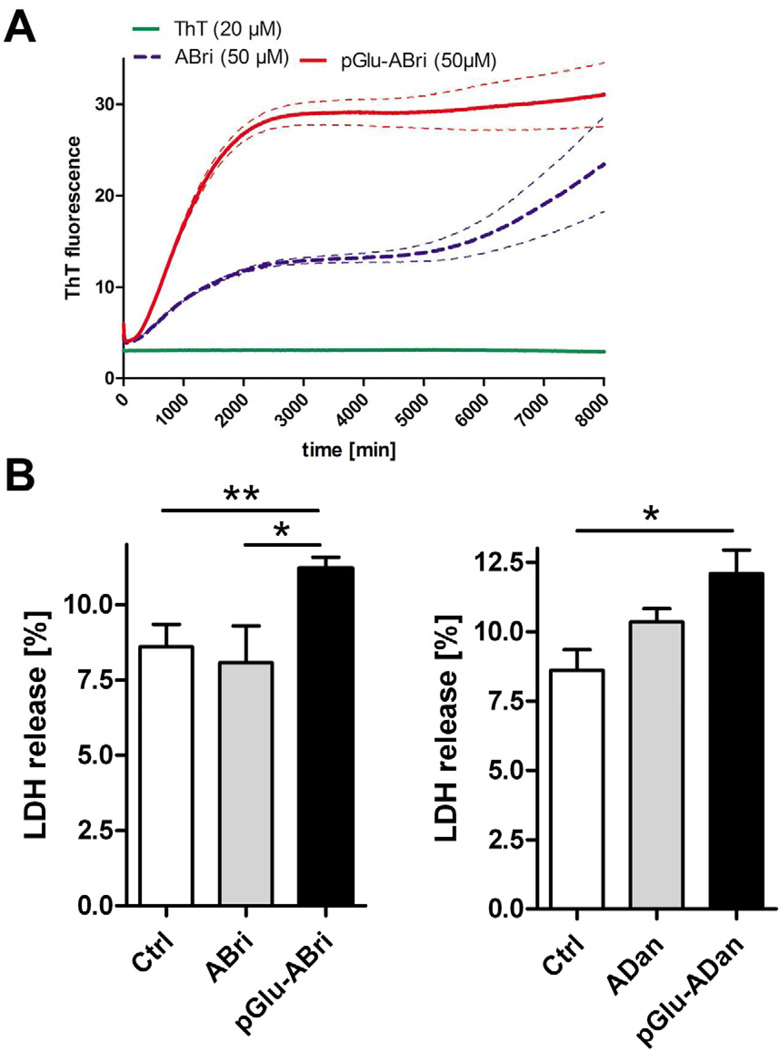
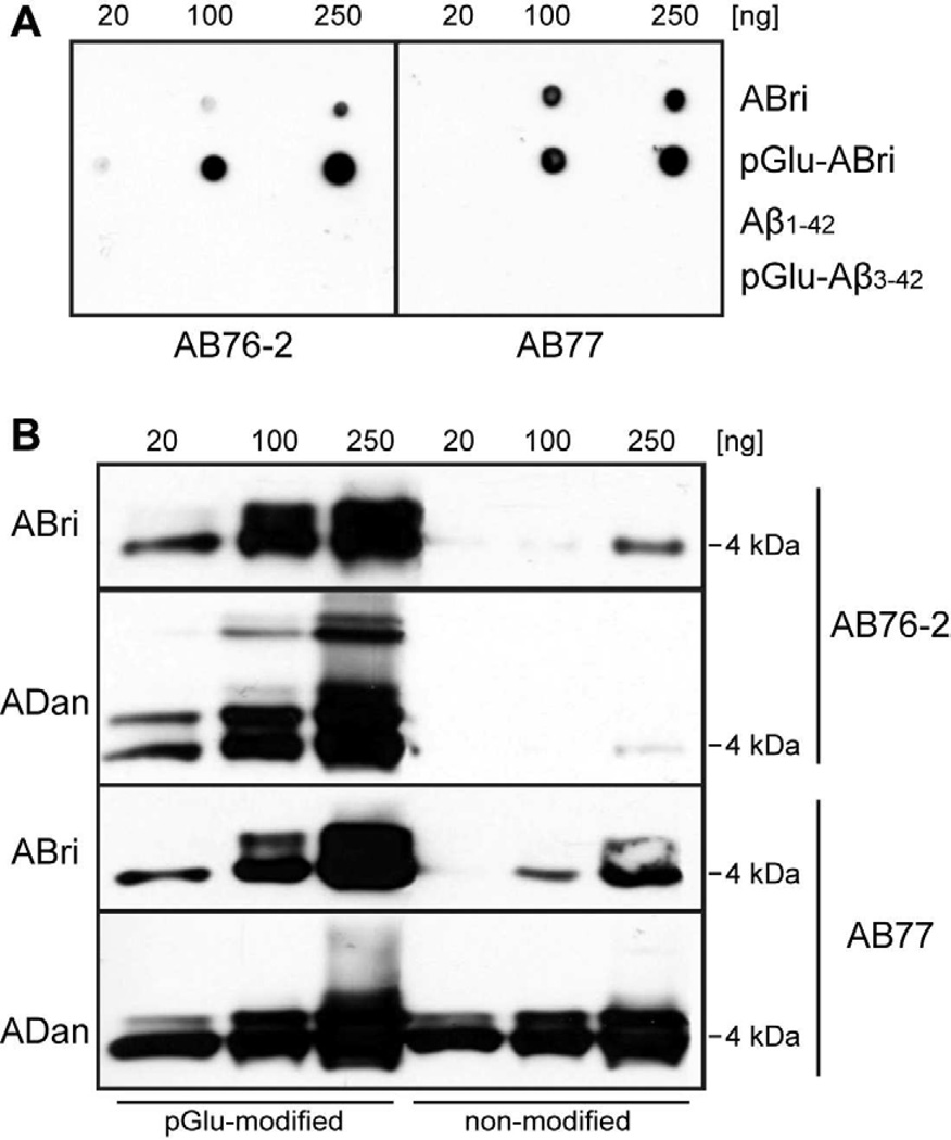
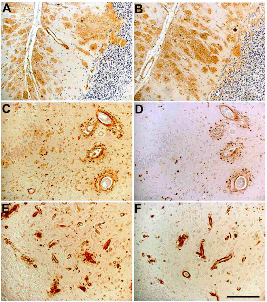
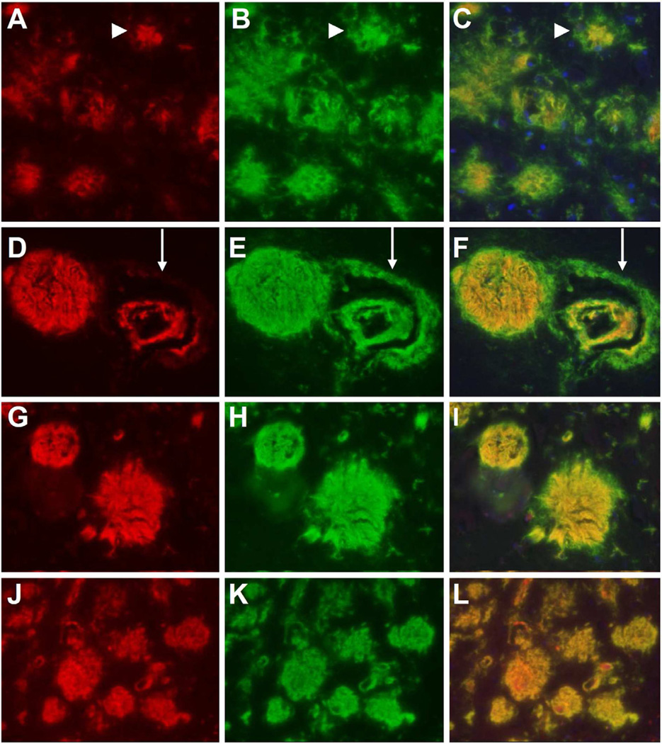
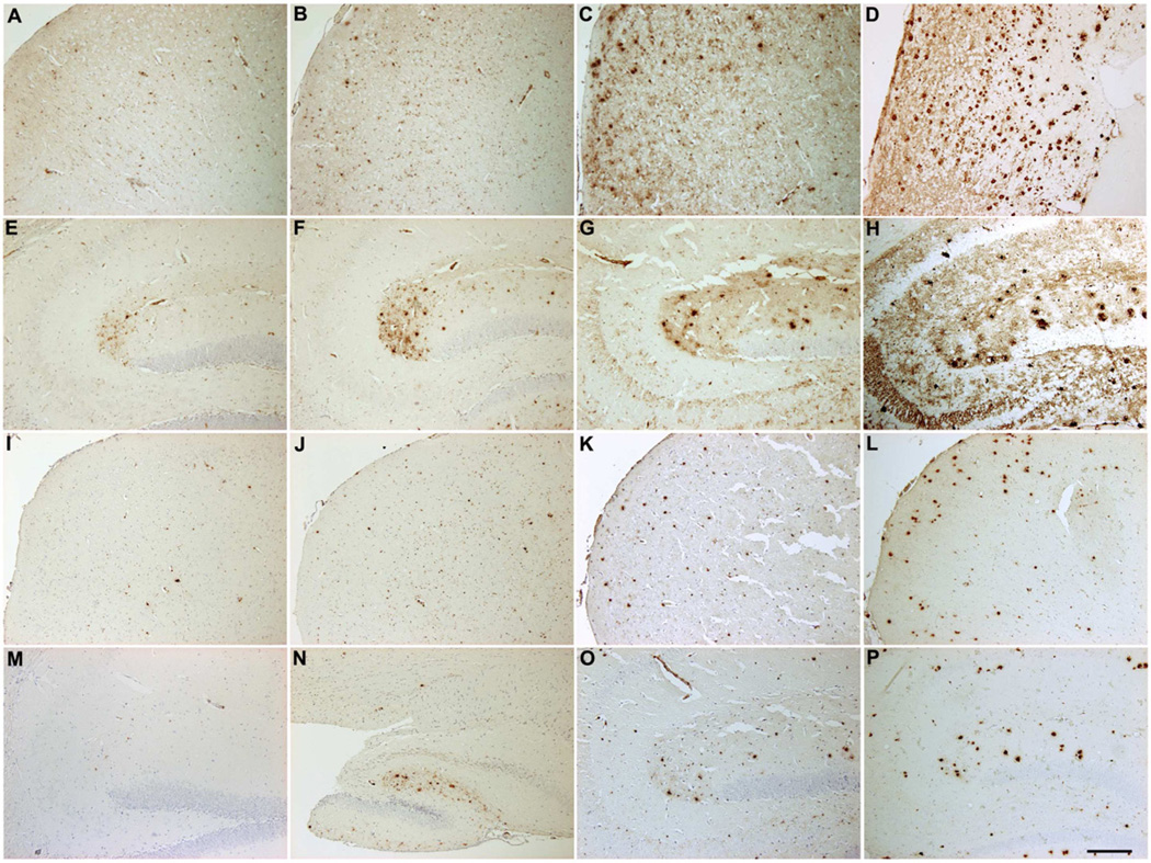
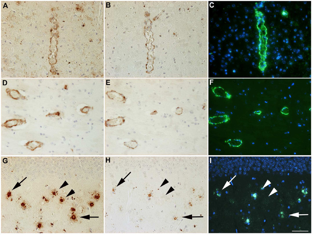
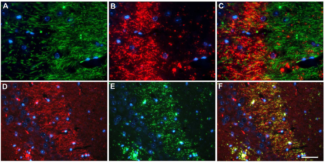
Familial British and familial Danish dementia (FDD) are progressive neurodegenerative disorders characterized by cerebral deposition of the amyloidogenic peptides ABri and ADan, respectively. These amyloid peptides start with an N-terminal glutamate residue, which can be posttranslationally converted into a pyroglutamate (pGlu) modified form, a mechanism which has been extensively described to be relevant for amyloid-beta (Aβ) peptides in Alzheimer's disease. Like pGlu-Aβ peptides, pGlu-ABri peptides have an increased aggregation propensity and show higher toxicity on human neuroblastoma cells as their nonmodified counterparts. We have generated novel N-terminal specific antibodies detecting the pGlu-modified forms of ABri and ADan peptides. With these antibodies we were able to identify abundant extracellular amyloid plaques, vascular, and parenchymal deposits in human familial British dementia and FDD brain tissue, and in a mouse model for FDD. Double-stainings using C-terminal specific antibodies in human samples revealed that highly aggregated pGlu-ABri and pGlu-ADan peptides are mainly present in plaque cores and central vascular deposits, leading to the assumption that these peptides have seeding properties. Furthermore, in an FDD-mouse model ADan peptides were detected in presynaptic terminals of the hippocampus where they might contribute to impaired synaptic transmission. These similarities of ABri and ADan to Aβ in Alzheimer's disease suggest that the posttranslational pGlu-modification of amyloid peptides might represent a general pathological mechanism leading to increased aggregation and toxicity in these forms of degenerative dementias.
Copyright © 2013 Elsevier Inc. All rights reserved.
Conflict of interest statement
The authors disclose no conflict of interest.
Animal studies were performed with the approval of the local research ethics committee in accordance with national and international guidelines.
Figures
Similar articles
-
Chemical traits of cerebral amyloid angiopathy in familial British-, Danish-, and non-Alzheimer's dementias.J Neurochem. 2022 Nov;163(3):233-246. doi: 10.1111/jnc.15694. Epub 2022 Sep 25. J Neurochem. 2022. PMID: 36102248 Free PMC article.
-
Amyloid peptides ABri and ADan show differential neurotoxicity in transgenic Drosophila models of familial British and Danish dementia.Mol Neurodegener. 2014 Jan 9;9:5. doi: 10.1186/1750-1326-9-5. Mol Neurodegener. 2014. PMID: 24405716 Free PMC article.
-
Pyroglutamate formation influences solubility and amyloidogenicity of amyloid peptides.Biochemistry. 2009 Jul 28;48(29):7072-8. doi: 10.1021/bi900818a. Biochemistry. 2009. PMID: 19518051
-
Chromosome 13 dementias.Cell Mol Life Sci. 2005 Aug;62(16):1814-25. doi: 10.1007/s00018-005-5092-5. Cell Mol Life Sci. 2005. PMID: 15968464 Free PMC article. Review.
-
Preamyloid lesions and cerebrovascular deposits in the mechanism of dementia: lessons from non-beta-amyloid cerebral amyloidosis.Neurodegener Dis. 2008;5(3-4):173-5. doi: 10.1159/000113694. Epub 2008 Mar 6. Neurodegener Dis. 2008. PMID: 18322382 Free PMC article. Review.
Cited by
-
Are N- and C-terminally truncated Aβ species key pathological triggers in Alzheimer's disease?J Biol Chem. 2018 Oct 5;293(40):15419-15428. doi: 10.1074/jbc.R118.003999. Epub 2018 Aug 24. J Biol Chem. 2018. PMID: 30143530 Free PMC article. Review.
-
Oxidative stress and mitochondria-mediated cell death mechanisms triggered by the familial Danish dementia ADan amyloid.Neurobiol Dis. 2016 Jan;85:130-143. doi: 10.1016/j.nbd.2015.10.003. Epub 2015 Oct 13. Neurobiol Dis. 2016. PMID: 26459115 Free PMC article.
-
Mitochondrial dysfunction induced by a post-translationally modified amyloid linked to a familial mutation in an alternative model of neurodegeneration.Biochim Biophys Acta. 2014 Dec;1842(12 Pt A):2457-67. doi: 10.1016/j.bbadis.2014.09.010. Epub 2014 Sep 28. Biochim Biophys Acta. 2014. PMID: 25261792 Free PMC article.
-
Chemical traits of cerebral amyloid angiopathy in familial British-, Danish-, and non-Alzheimer's dementias.J Neurochem. 2022 Nov;163(3):233-246. doi: 10.1111/jnc.15694. Epub 2022 Sep 25. J Neurochem. 2022. PMID: 36102248 Free PMC article.
-
Pyroglutamyl-N-terminal prion protein fragments in sheep brain following the development of transmissible spongiform encephalopathies.Front Mol Biosci. 2015 Mar 11;2:7. doi: 10.3389/fmolb.2015.00007. eCollection 2015. Front Mol Biosci. 2015. PMID: 25988175 Free PMC article.
References
-
- Alexandru A, Jagla W, Graubner S, Becker A, Bauscher C, Kohlmann S, Sedlmeier R, Raber KA, Cynis H, Ronicke R, Reymann KG, Petrasch-Parwez E, Hartlage-Rubsamen M, Waniek A, Rossner S, Schilling S, Osmand AP, Demuth HU, von Horsten S. Selective Hippocampal Neurodegeneration in Transgenic Mice Expressing Small Amounts of Truncated A{beta} Is Induced by Pyroglutamate-A{beta} Formation. J Neurosci. 2011;31(36):12790–12801. - PMC - PubMed
-
- Benilova I, Karran E, De Strooper B. The toxic Abeta oligomer and Alzheimer's disease: an emperor in need of clothes. Nat Neurosci. 2012;15(3):349–357. - PubMed
-
- Busby WH, Jr, Quackenbush GE, Humm J, Youngblood WW, Kizer JS. An enzyme(s) that converts glutaminyl-peptides into pyroglutamyl-peptides. Presence in pituitary, brain, adrenal medulla, and lymphocytes. J Biol Chem. 1987;262(18):8532–8536. - PubMed
-
- Casas C, Sergeant N, Itier JM, Blanchard V, Wirths O, van der Kolk N, Vingtdeux V, van de Steeg E, Ret G, Canton T, Drobecq H, Clark A, Bonici B, Delacourte A, Benavides J, Schmitz C, Tremp G, Bayer TA, Benoit P, Pradier L. Massive CA1/2 neuronal loss with intraneuronal and N-terminal truncated Abeta42 accumulation in a novel Alzheimer transgenic model. Am J Pathol. 2004;165(4):1289–1300. - PMC - PubMed
-
- Christensen DZ, Bayer TA, Wirths O. Intracellular Abeta triggers neuron loss in the cholinergic system of the APP/PS1KI mouse model of Alzheimer's disease. Neurobiol Aging. 2010;31(7):1153–1163. - PubMed
Publication types
MeSH terms
Substances
Grants and funding
LinkOut - more resources
Full Text Sources
Other Literature Sources
Medical
Miscellaneous
