

Higher susceptibility to experimental autoimmune encephalomyelitis in Muc1-deficient mice is associated with increased Th1/Th17 responses
- PMID: 23261777
- PMCID: PMC3587144
- DOI: 10.1016/j.bbi.2012.12.004
Higher susceptibility to experimental autoimmune encephalomyelitis in Muc1-deficient mice is associated with increased Th1/Th17 responses
Abstract
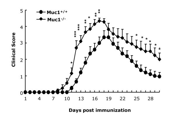
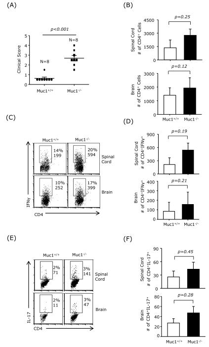
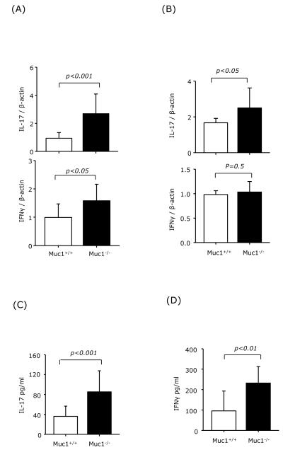
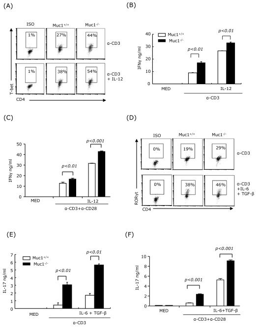
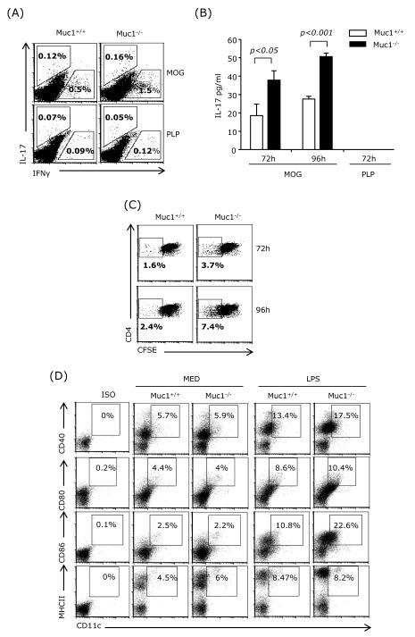
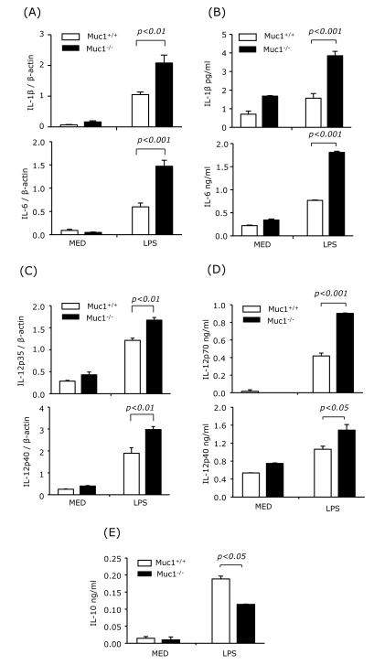
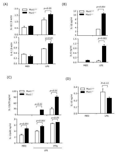
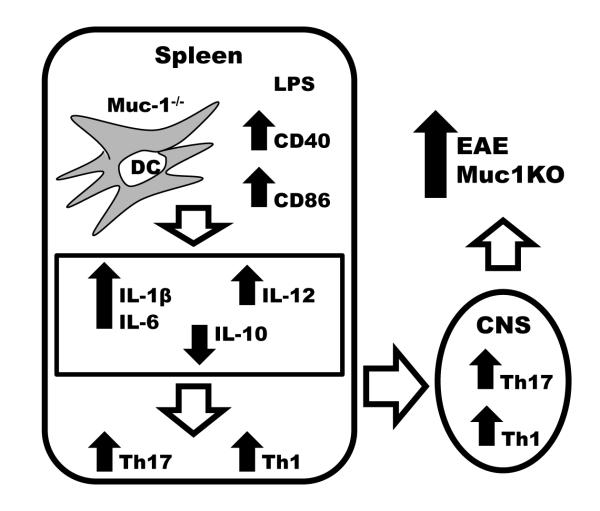
Multiple sclerosis (MS) is an autoimmune disease of the central nervous system in which dendritic cells (DC) play an important role in the development of inflammatory responses. Recently it has been shown that Muc1, a membrane tethered glycoprotein, has an ability to suppress inflammatory responses in cultured DC. The objective of this study was to investigate the possible involvement of Muc1 in the development of MS using experimental autoimmune encephalomyelitis (EAE) in mice, a widely used animal model of MS. Our results showed that: (1) Muc1(-/-) mice developed greater EAE severity compared with wild type (wt) mice, which correlated with increased numbers of Th1 and Th17 cells infiltrating into the CNS; (2) upon stimulation, splenic DC from Muc1(-/-) mice produced greater amounts of IL-1β, IL-6, and IL-12 but less amounts of IL-10 compared with those from wt mice; and (3) the ability of splenic DC to differentiate antigen-specific CD4+ T cells into Th1 and Th17 cells was greater in Muc1(-/-) mice compared with wt mice. We conclude that Muc1 plays an anti-inflammatory role in EAE. This is the first report demonstrating the possible involvement of Muc1 in the development of MS and might provide a potential target for immunotherapy.
Copyright © 2012 Elsevier Inc. All rights reserved.
Figures

Similar articles
-
IL-7/IL-7 Receptor Signaling Differentially Affects Effector CD4+ T Cell Subsets Involved in Experimental Autoimmune Encephalomyelitis.J Immunol. 2015 Sep 1;195(5):1974-83. doi: 10.4049/jimmunol.1403135. Epub 2015 Jul 29. J Immunol. 2015. PMID: 26223651 Free PMC article.
-
Role of Th17 cells in the pathogenesis of CNS inflammatory demyelination.J Neurol Sci. 2013 Oct 15;333(1-2):76-87. doi: 10.1016/j.jns.2013.03.002. Epub 2013 Apr 8. J Neurol Sci. 2013. PMID: 23578791 Free PMC article. Review.
-
MicroRNA223 promotes pathogenic T-cell development and autoimmune inflammation in central nervous system in mice.Immunology. 2016 Aug;148(4):326-38. doi: 10.1111/imm.12611. Epub 2016 Jun 29. Immunology. 2016. PMID: 27083389 Free PMC article.
-
Infiltration of Th1 and Th17 cells and activation of microglia in the CNS during the course of experimental autoimmune encephalomyelitis.Brain Behav Immun. 2010 May;24(4):641-51. doi: 10.1016/j.bbi.2010.01.014. Epub 2010 Feb 6. Brain Behav Immun. 2010. PMID: 20138983
-
Th17 Cells in MS and Experimental Autoimmune Encephalomyelitis.Int MS J. 2009 Apr;16(1):12-8. Int MS J. 2009. PMID: 19413921 Review.
Cited by
-
The Role of MUC1 in Renal Cell Carcinoma.Biomolecules. 2024 Mar 7;14(3):315. doi: 10.3390/biom14030315. Biomolecules. 2024. PMID: 38540735 Free PMC article. Review.
-
Chimeric antigen receptor natural killer cells: a promising antitumor immunotherapy.MedComm (2020). 2023 Dec 1;4(6):e422. doi: 10.1002/mco2.422. eCollection 2023 Dec. MedComm (2020). 2023. PMID: 38045827 Free PMC article. Review.
-
Shared genetic effect of kidney function on bipolar and major depressive disorders: a large-scale genome-wide cross-trait analysis.Hum Genomics. 2024 Jun 11;18(1):60. doi: 10.1186/s40246-024-00627-3. Hum Genomics. 2024. PMID: 38858783 Free PMC article.
-
Serum carbohydrate antigen 153 and renal function in patients with type 2 diabetes mellitus.J Clin Lab Anal. 2018 Sep;32(7):e22461. doi: 10.1002/jcla.22461. Epub 2018 Apr 27. J Clin Lab Anal. 2018. PMID: 29701319 Free PMC article.
-
MUC1 Mucin: A Putative Regulatory (Checkpoint) Molecule of T Cells.Front Immunol. 2018 Oct 22;9:2391. doi: 10.3389/fimmu.2018.02391. eCollection 2018. Front Immunol. 2018. PMID: 30405607 Free PMC article. Review.
References
-
- Agrawal B, Krantz MJ, Parker J, Longenecker BM. Expression of MUC1 mucin on activated human T cells: implications for a role of MUC1 in normal immune regulation. Cancer Res. 1998;58:4079–4081. - PubMed
-
- Auriemma M, Brzoska T, Klenner L, Kupas V, Goerge T, Voskort M, Zhao Z, Sparwasser T, Luger TA, Loser K. alpha-MSH-Stimulated Tolerogenic Dendritic Cells Induce Functional Regulatory T Cells and Ameliorate Ongoing Skin Inflammation. The Journal of investigative dermatology. 2012;132:1814–1824. - PubMed
-
- Bailey SL, Schreiner B, McMahon EJ, Miller SD. CNS myeloid DCs presenting endogenous myelin peptides ‘preferentially’ polarize CD4+ T(H)-17 cells in relapsing EAE. Nature immunology. 2007;8:172–180. - PubMed
Publication types
MeSH terms
Substances
Grants and funding
LinkOut - more resources
Full Text Sources
Other Literature Sources
Research Materials
Miscellaneous
