. 2013 Feb 15;112(4):675-88.
doi: 10.1161/CIRCRESAHA.111.300502. Epub 2012 Dec 20.

Matrix metalloproteinase-28 deletion exacerbates cardiac dysfunction and rupture after myocardial infarction in mice by inhibiting M2 macrophage activation

Affiliations

Matrix metalloproteinase-28 deletion exacerbates cardiac dysfunction and rupture after myocardial infarction in mice by inhibiting M2 macrophage activation

Yonggang Ma et al. Circ Res. .

Abstract

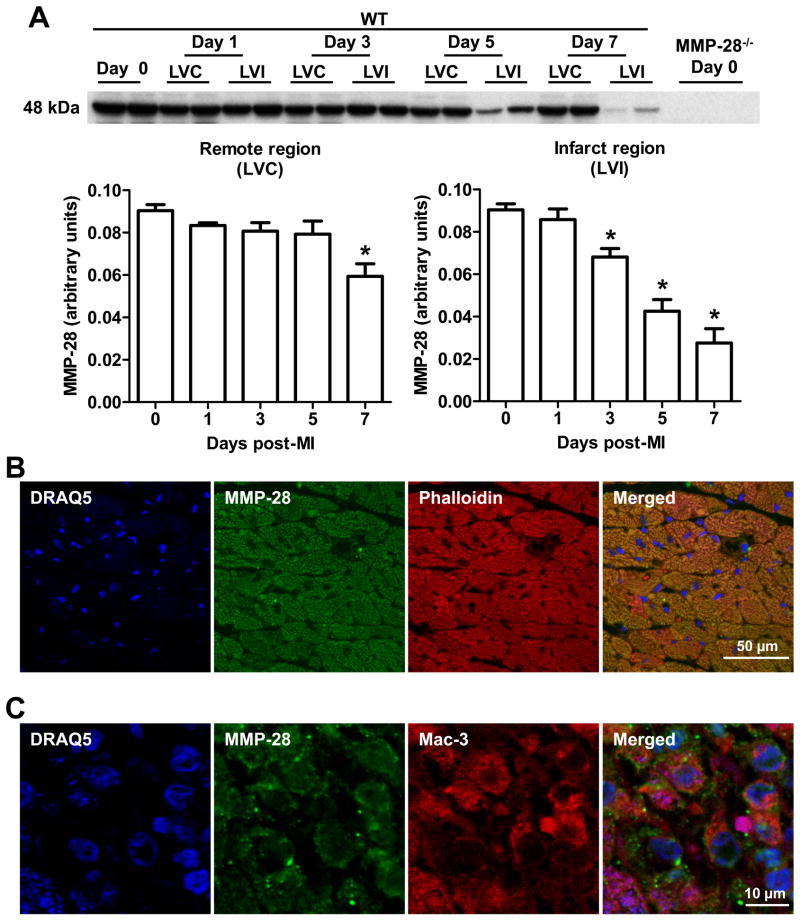
Rationale: Matrix metalloproteinase (MMP)-28 regulates the inflammatory and extracellular matrix responses in cardiac aging, but the roles of MMP-28 after myocardial infarction (MI) have not been explored.

Objective: To determine the impact of MMP-28 deletion on post-MI remodeling of the left ventricle (LV).

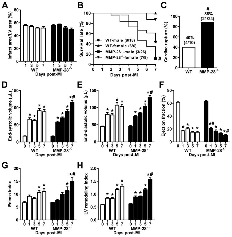
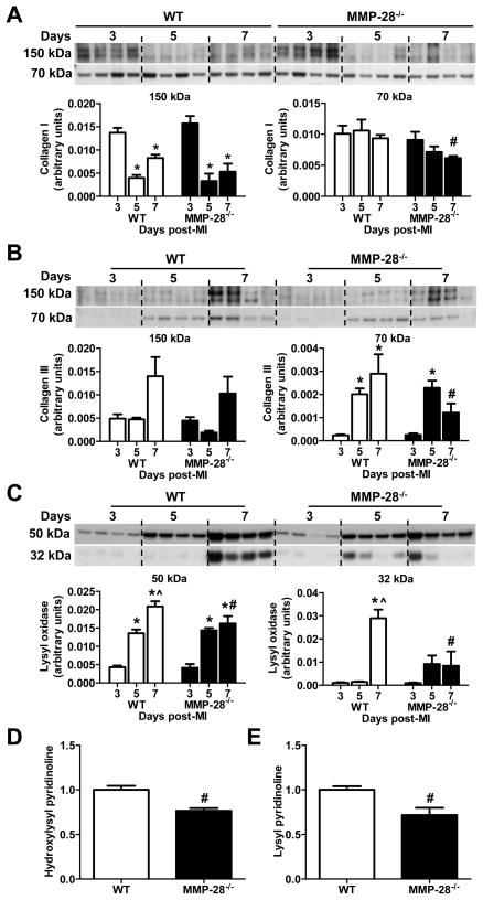
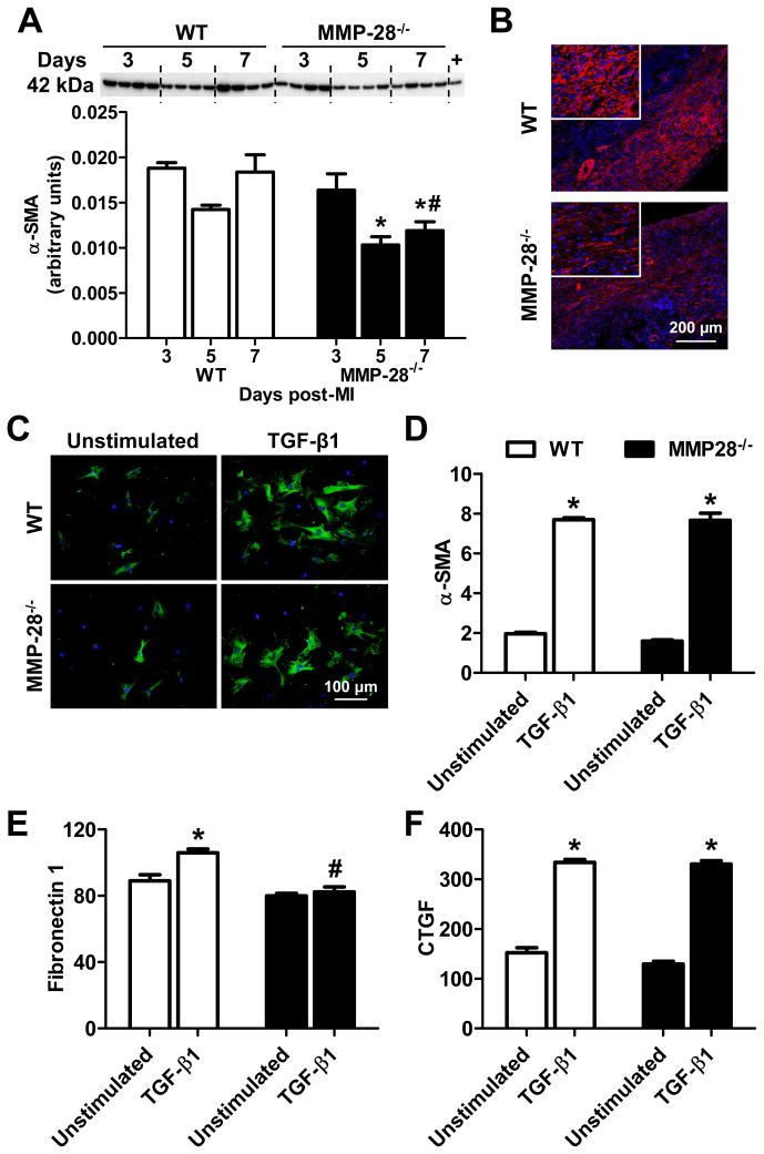
Methods and results: Adult C57BL/6J wild-type (n=76) and MMP null (MMP-28((-/-)), n=86) mice of both sexes were subjected to permanent coronary artery ligation to create MI. MMP-28 expression decreased post-MI, and its cell source shifted from myocytes to macrophages. MMP-28 deletion increased day 7 mortality because of increased cardiac rupture post-MI. MMP-28(-/-) mice exhibited larger LV volumes, worse LV dysfunction, a worse LV remodeling index, and increased lung edema. Plasma MMP-9 levels were unchanged in the MMP-28((-/-)) mice but increased in wild-type mice at day 7 post-MI. The mRNA levels of inflammatory and extracellular matrix proteins were attenuated in the infarct regions of MMP-28(-/-) mice, indicating reduced inflammatory and extracellular matrix responses. M2 macrophage activation was impaired when MMP-28 was absent. MMP-28 deletion also led to decreased collagen deposition and fewer myofibroblasts. Collagen cross-linking was impaired as a result of decreased expression and activation of lysyl oxidase in the infarcts of MMP-28(-/-) mice. The LV tensile strength at day 3 post-MI, however, was similar between the 2 genotypes.

Conclusions: MMP-28 deletion aggravated MI-induced LV dysfunction and rupture as a result of defective inflammatory response and scar formation by suppressing M2 macrophage activation.


Figures

Figure 1
Figure 1. Expression patterns and cellular source of MMP-28 at baseline and post-MI
A, MMP-28 expression decreased in both remote and infarct regions post-MI. MMP-28−/− day 0 hearts were used as negative controls. n=4–6/group. *p<0.05 vs. day 0. B, MMP-28 co-localized with phalloidin, a marker of myocytes, at baseline (day 0). C, MMP-28 from macrophages increased in the infarct region post-MI. MMP-28 co-localized with Mac-3, a marker of macrophages, at day 5 post-MI. Representative images from n=3 stained LVs for B and C. Blue=DRAQ5, Green=MMP-28, Red=phalloidin or Mac-3.
Figure 2
Figure 2. MMP-28 deletion decreased survival, increased cardiac rupture, and exacerbated LV dysfunction post-MI
A, Infarct areas were similar between WT and MMP-28−/− mice. n=8–14/group. B, MMP-28 deletion reduced 7 day post-MI survival. n=6–26/group. C, Cardiac rupture post-MI was increased in MMP-28−/− mice. n= 10–24/group. D and E, LV end systolic and diastolic volumes at day 7 post-MI were higher in MMP-28−/−, compared with WT. F, Ejection fraction in MMP-28−/− mice was attenuated at day 1, but exacerbated at day 7 post-MI, compared with WT. G, The ratio of lung weight to tibia length (edema index) at day 7 post-MI was higher in MMP-28−/− than in WT. H, MMP-28 deletion showed higher EDV/ LV mass (LV remodeling index) at day 7 post-MI. n=8–14/group for D through H. *p<0.05 vs. day 0 and #p<0.05 vs. WT.
Figure 3
Figure 3. M2 macrophage activation was impaired with MMP-28 deletion
A, Galectin-3 expression was similarly induced in WT and MMP-28−/− mice post-MI. Isolated peritoneal macrophages were used as positive controls. n=4/group. B, Macrophage numbers infiltrated into infarct area at day 5 post-MI showed no difference between WT and MMP-28−/− mice. n=5–7/group. C, MMP-28 deletion did not affect M1 macrophage markers (except IL-1β) in day 7 infarct. D, MMP-28 deletion attenuated M2 macrophage markers in day 7 infarct. n=6/group for C and D. *p<0.05 vs. day 3 and #p<0.05 vs. WT.
Figure 4
Figure 4. MMP-28 deletion attenuated macrophages polarization towards M2 subtype
Peritoneal macrophages were stimulated with IFN-γ+LPS or IL-4. A, MMP-28 expression was similar among different macrophage subtypes. B, MMP-28 deletion did not affect macrophage activation towards M1 subtype. C, MMP-28 deletion attenuated macrophage polarization towards M2 phenotype. n=4/group for A through C. *p<0.05 vs. unstimulated and #p<0.05 vs. WT.
Figure 5
Figure 5. Collagen deposition, lysyl oxidase content, and collagen cross-linking were reduced with MMP-28 deletion
A and B, MMP-28 deletion led to reduced collagen I and collagen III accumulation at day 7 post-MI. C, Both precursor and active forms of lysyl oxidase at day 7 post-MI were suppressed with MMP-28 deletion. n=4/group for A through C. Dand E, Hydroxylysyl pyridinoline and lysyl pyridinoline in day 7 infarct were reduced with MMP-28 deletion. n=3/group. *p<0.05 vs. day 3, ^p<0.05 vs. day 5, and #p<0.05 vs. WT day 7.
Figure 6
Figure 6. MMP-28 deletion reduced myofibroblast numbers at day 7 post-MI and affected myofibroblast function
A, α-SMA expression at day 7 post-MI was reduced in MMP-28−/− mice, compared to WT. Isolated cardiac fibroblasts were used as positive controls. n=4/group. B, Immunofluorescence staining showed that MMP-28−/− mice had fewer myofibroblast numbers in the non-vessel interstitial area at day 7 after MI. Blue=DAPI, red=α-SMA. Representative images from n=3 stained LVs. C and D, TGF-β1 induced similar upregulation in α-SMA in both WT and MMP-28−/−cardiac fibroblasts. E, Fibronectin 1 was lower in MMP-28−/− fibroblasts stimulated with TGF-β1, compared to WT. F, TGF-β1 induced similar upregulation in CTGF in both genotypes. n=4/group for C through F. *p<0.05 vs. unstimulated and #p<0.05 vs. WT.

Comment in

References

    1. Lambert JM, Lopez EF, Lindsey ML. Macrophage roles following myocardial infarction. Int J Cardiol. 2008;130:147–158. - PMC - PubMed
    1. Frangogiannis NG. Regulation of the inflammatory response in cardiac repair. Circ Res. 2012;110:159–173. - PMC - PubMed
    1. Nahrendorf M, Pittet MJ, Swirski FK. Monocytes: Protagonists of infarct inflammation and repair after myocardial infarction. Circulation. 2010;121:2437–2445. - PMC - PubMed
    1. Ma Y, Lindsey ML, Halade GV. Dha derivatives of fish oil as dietary supplements: A nutrition-based drug discovery approach for therapies to prevent metabolic cardiotoxicity. Expert Opin Drug Discov. 2012 - PMC - PubMed
    1. Matsui Y, Ikesue M, Danzaki K, Morimoto J, Sato M, Tanaka S, Kojima T, Tsutsui H, Uede T. Syndecan-4 prevents cardiac rupture and dysfunction after myocardial infarction. Circ Res. 2011;108:1328–1339. - PubMed

Publication types

MeSH terms