. 2013 Feb 8;288(6):4475-85.
doi: 10.1074/jbc.M112.436386. Epub 2012 Dec 23.

Interactions of Anopheles gambiae odorant-binding proteins with a human-derived repellent: implications for the mode of action of n,n-diethyl-3-methylbenzamide (DEET)

Affiliations

Interactions of Anopheles gambiae odorant-binding proteins with a human-derived repellent: implications for the mode of action of n,n-diethyl-3-methylbenzamide (DEET)

Emma J Murphy et al. J Biol Chem. .

Abstract

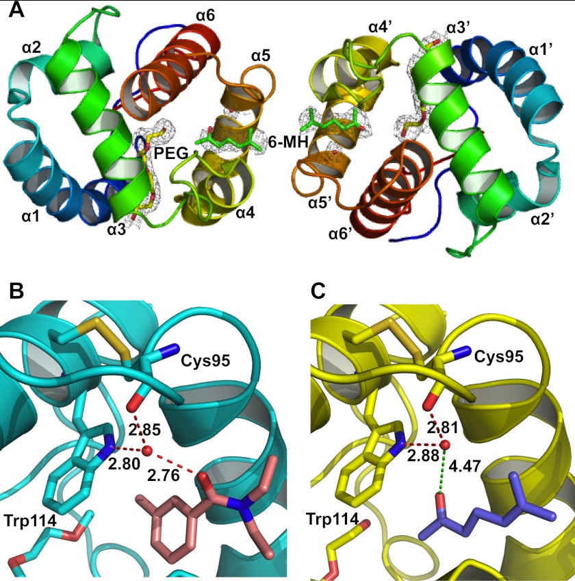
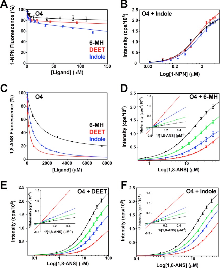
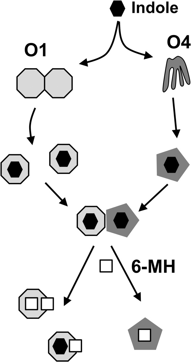
The Anopheles gambiae mosquito, which is the vector for Plasmodium falciparum malaria, uses a series of olfactory cues emanating from human sweat to select humans as their source for a blood meal. Perception of these odors within the mosquito olfactory system involves the interplay of odorant-binding proteins (OBPs) and odorant receptors and disrupting the normal responses to those odorants that guide mosquito-human interactions represents an attractive approach to prevent the transmission of malaria. Previously, it has been shown that DEET targets multiple components of the olfactory system, including OBPs and odorant receptors. Here, we present the crystal structure of A. gambiae OBP1 (OBP1) in the complex it forms with a natural repellent 6-methyl-5-heptene-2-one (6-MH). We find that 6-MH binds to OBP1 at exactly the same site as DEET. However, key interactions with a highly conserved water molecule that are proposed to be important for DEET binding are not involved in binding of 6-MH. We show that 6-MH and DEET can compete for the binding of attractive odorants and in doing so disrupt the interaction that OBP1 makes with OBP4. We further show that 6-MH and DEET can bind simultaneously to OBPs with other ligands. These results suggest that the successful discovery of novel reagents targeting OBP function requires knowledge about the specific mechanism of binding to the OBP rather than their binding affinity.


Figures

FIGURE 1.
FIGURE 1.
Crystal structure of the OBP1–6-MH complex. A, schematic representation of the overall structure of the OBP1 dimer showing two molecules of 6-MH (green) bound at the interface and two molecules of PEG (yellow) filling the rest of the binding pocket. The density from a 2FoFc map is contoured at 1 σ in gray, and waters are shown in red. B, schematic representation of the OBP1-DEET complex. Hydrogen bonds between DEET (salmon), Trp-114, Cys-95, and a water molecule are shown in red and labeled with interatomic distances (Å). C, schematic representation of the OBP1–6-MH complex. The carbonyl group of 6-MH (blue) is 4.47 Å away from the conserved water molecule and so is not within hydrogen bonding distance. The water molecule that is conserved in all three OBP structures published to date is shown in red.
FIGURE 2.
FIGURE 2.
There is no interaction between OBP1 and 4 in the presence of 6-MH. A, 1H-15N HSQC spectrum of 15N-OBP1 (O1) in the presence of 3 mm 6-MH (black) overlaid with the spectrum recorded in the presence of OBP4 (O4) (red). The spectra are essentially identical, consistent with a lack of interaction between OBP1 and OBP4. B, 1H-15N HSQC spectrum of 15N-OBP4 with 3 mm 6-MH (black). Addition of OBP1 (red) does not result in any chemical shift changes confirming the lack of interaction between the two proteins in the presence of 6-MH. C, 6-MH competing with indole for binding to OBP1 in the presence of OBP4. Spectrum of 15N-OBP1 plus indole plus OBP4 (black) overlaid with the spectrum of 15N-OBP1 plus indole plus OBP4 plus 6-MH (red) is shown. The black arrows indicate peaks only observed when OBP1 is bound to 6-MH. D, DEET inducing conformational ordering of OBP4 but not allowing interaction between OBP1 and 4. 1H-15N HSQC spectrum of OBP4 in the presence of DEET (black) overlaid with the spectrum of OBP4 plus DEET plus OBP1 (red) is shown. The lack of shifts indicates no interactions between the two proteins. All spectra were recorded in 20 mm sodium phosphate, pH 7.4, at 25 °C.
FIGURE 3.
FIGURE 3.
15N NMR relaxation measurements for the OBP1–6MH complex. 15N T1 (longitudinal) relaxation times and 15N T2 (transverse) relaxation times as a function of residue number are shown. The locations of the elements of secondary structure are shown at the top. Dashed lines represent the mean values of T1 = 744.5 ± 34.7 ms and T2 = 72.6 ± 6.7 ms. Values for Thr-3, Asp-48, Lys-50, and Ala-88 were excluded from these calculations because their T2 values deviated significantly from the mean. Spectra were recorded in 20 mm sodium phosphate, pH 7.4, at 25 °C. Assignments were extrapolated from those made with OBP1-indole; only unambiguous peaks were assigned.
FIGURE 4.
FIGURE 4.
Assay of OBP1 binding to 1-NPN in the presence of 6-MH, DEET, indole, and citronellal. A, competition binding assay. OBP1 (O1) in the presence of 1-NPN (both 1 μm in 50 mm Tris-HCl, pH 7.4) was titrated with 6-MH (black), DEET (red), indole (blue), or citronellal (green) to final concentrations of ∼200 μm. Fluorescence intensity is normalized to the value in the absence of ligand. B, heterologous binding assay revealing that 6-MH does not compete with 1-NPN binding. OBP1 (1 μm) in the absence (black) or presence of 6-MH (50 μm, blue; 200 μm, red) was titrated with 1-NPN to a final concentration of 13 μm. C, citronellal competing for binding of 1-NPN. OBP1 in the absence (black) or presence of citronellal (5 μm, red; 10 μm, blue; 50 μm, green; or 100 μm, orange) was titrated with 1-NPN to a final concentration of 11 μm. D, DEET and indole not competing with 1-NPN for OBP1 binding. Log plots of OBP1 titrated with 1-NPN to a final concentration of 13.5 μm, in the absence (black) or presence of DEET (20 μm, red; 100 μm, blue) are shown. E, as in D with indole as opposed to DEET. 1-NPN and ligand solutions were in methanol, and all curves are an average of three replicates, and error bars are shown. In B--E, OBP1 was 1 μm in 50 mm Tris-HCl, pH 7.4, and the concentration of 1-NPN was 1 mm in methanol.
FIGURE 5.
FIGURE 5.
Assay of 6-MH, DEET, and indole binding to OBP4. A, 6-MH, DEET, and indole do not displace 1-NPN from OBP4 (O4). OBP4 in the presence of 1-NPN (both 0. 5 μm in 50 mm Tris-HCl, pH 7.4) was titrated with a 10 mm 6-MH (black), 10 mm DEET (red), or 10 mm indole (blue) to final concentrations of ≈200 μm. Fluorescence intensity is normalized to the value in the absence of ligand. B, indole does not compete for 1-NPN binding: OBP4 (0.5 μm) in the absence (black) or presence of indole (5 μm, blue; 50 μm, red) was titrated with 1-NPN (1 mm) to a final concentration of 6 μm. C, increasing concentrations of 6-MH (black), DEET (red), and indole (blue) can displace 1,8-ANS (20 μm) from OBP4 (5 μm). Fluorescence intensity is displayed as percentage of the value in the absence of ligand. D–F, log plots of 1,8-ANS titrations into OBP4 (5 μm in 50 mm Tris-HCl, pH 7.4) is shown in the absence (black) or presence of (D) 6-MH, 500 μm (green), 1000 μm (blue), or 5000 μm (red); (E) DEET, 100 μm (green), 200 μm (blue), or 500 μm (red); (F) indole, 235 μm (green), 470 μm (blue), or 1088 μm (red). The Lineweaver-Burk plots (insets in D–F) are linear and intercept the y axis at the same point showing competitive binding in each case. All ligands were added as solutions in methanol to a maximum methanol concentration of 1%. Each curve is the average of three replicates and error bars are shown; these are within the limits of the symbol for the Lineweaver-Burk plots.
FIGURE 6.
FIGURE 6.
Scheme of ligand and repellent binding to OBP1 and 4. In the absence of ligands OBP1 (light gray octagon) appears to exist predominantly as dimers, whereas OBP4 (dark gray) is a highly dynamic structure. Binding of odorants, including indole (black), leads to dissociation of OBP1 into monomers and a stabilization of the OBP4 structure that allows the two proteins to form heterodimers. Repellent molecules, like 6-MH (white square) can function either to compete for and displace the normal ligand or can bind at the same time as the native ligand and disrupt the formation of the OBP1-OBP4 complex.

References

    1. Crawford J. E., Lazzaro B. P. (2010) The demographic histories of the M and S molecular forms of Anopheles gambiae s.s. Mol. Biol. Evol. 27, 1739–1744 - PMC - PubMed
    1. Dondorp A. M., Nosten F., Yi P., Das D., Phyo A. P., Tarning J., Lwin K. M., Ariey F., Hanpithakpong W., Lee S. J., Ringwald P., Silamut K., Imwong M., Chotivanich K., Lim P., Herdman T., An S. S., Yeung S., Singhasivanon P., Day N. P., Lindegardh N., Socheat D., White N. J. (2009) Artemisinin resistance in Plasmodium falciparum malaria. New Engl. J. Med. 361, 455–467 - PMC - PubMed
    1. RTSS Clinical Trials Partnership (2012) A phase 3 trial of RTS,S/AS01 malaria vaccine in African infants. New Engl. J. Med. 367, 2284–2295 - PMC - PubMed
    1. Trape J. F., Rogier C. (1996) Combating malaria morbidity and mortality by reducing transmission. Parasitol. Today 12, 236–240 - PubMed
    1. Hallem E. A., Nicole Fox A., Zwiebel L. J., Carlson J. R. (2004) Olfaction: mosquito receptor for human-sweat odorant. Nature 427, 212–213 - PubMed

Publication types

MeSH terms

Associated data