

Review
doi: 10.1016/j.yjmcc.2012.12.009.
Epub 2012 Dec 20.
Cardiac phenotype of Duchenne Muscular Dystrophy: insights from cellular studies
Affiliations
- PMID: 23261966
- PMCID: PMC3615054
- DOI: 10.1016/j.yjmcc.2012.12.009
Item in Clipboard
Review
Cardiac phenotype of Duchenne Muscular Dystrophy: insights from cellular studies
J Mol Cell Cardiol.
2013 May.
Abstract
Dilated cardiomyopathy is a serious and almost inevitable complication of Duchenne Muscular Dystrophy, a devastating and fatal disease of skeletal muscle resulting from the lack of functional dystrophin, a protein linking the cytoskeleton to the extracellular matrix. Ultimately, it leads to congestive heart failure and arrhythmias resulting from both cardiac muscle fibrosis and impaired function of the remaining cardiomyocytes. Here we summarize findings obtained in several laboratories, focusing on cellular mechanisms that result in degradation of cardiac functions in dystrophy.
Copyright © 2012 Elsevier Ltd. All rights reserved.
Figures
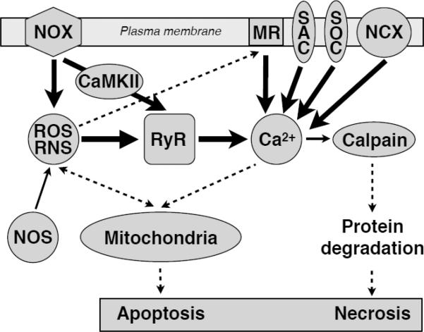
The abnormally high activity of several signaling pathways, such as ROS/RNS, hyperphosphorylation and Ca2+ signals, triggers mitochondrial damage pathways which ultimately may culminate in apoptotic or necrotic cell death.
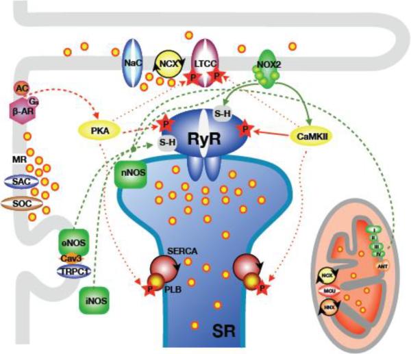
The diagram shows the main building blocks of cardiac Ca2+ signaling and EC-coupling. This is combined with the sources, pathways and targets of the pathomechanisms relevant for alterations of Ca2+ signaling in dystrophic cardiomyopathy. Green lines: pathways of oxidative and nitrosative signals (ROS/RNS). Red lines: pathways of protein phosphorylation signals. Solid lines depict pivotal and dashed lines depict secondary pathways. Dotted lines indicate additional targets of phosphorylation. Abbreviations not explained in the review: Cav3: caveolin-3; NaC: voltage-dependent Na+ channel. LTCC: L-type Ca2+ channel. AC: adenylate cyclase; MR: membrane ruptures; HNX: mitochondrial proton-sodium exchanger; MCU: mitochondrial Ca2+ uniporter; ANT: mitochondrial translocase.
References
-
- Clarac F, Massion J, Smith AM. Duchenne, Charcot and Babinski, three neurologists of La Salpetrière Hospital, and their contribution to concepts of the central organization of motor synergy. J Physiol Paris. 2009;103:361–76. - PubMed
-
- Finsterer J, Stöllberger C. The heart in human dystrophinopathies. Cardiology. 2003;99:1–19. - PubMed
-
- Hermans MCE, Pinto YM, Merkies ISJ, de Die-Smulders CEM, Crijns HJGM, Faber CG. Hereditary muscular dystrophies and the heart. Neuromuscul Disord. 2010;20:479–92. - PubMed
-
- Yilmaz A, Sechtem U. Cardiac involvement in muscular dystrophy: advances in diagnosis and therapy. Heart. 2012;98:420–9. - PubMed
-
- Aartsma-Rus A, Van Deutekom JCT, Fokkema IF, Van Ommen G-JB, Dunnen Den JT. Entries in the Leiden Duchenne muscular dystrophy mutation database: An overview of mutation types and paradoxical cases that confirm the reading-frame rule. Muscle Nerve. 2006;34:135–44. - PubMed
Publication types
MeSH terms
Substances
Grants and funding
LinkOut - more resources
Full Text Sources
Other Literature Sources
