. 2013 Jan 31;493(7434):679-83.
doi: 10.1038/nature11745. Epub 2012 Dec 23.

Regulation of mTORC1 by the Rag GTPases is necessary for neonatal autophagy and survival

Affiliations

Regulation of mTORC1 by the Rag GTPases is necessary for neonatal autophagy and survival

Alejo Efeyan et al. Nature. .

Abstract

The mechanistic target of rapamycin complex 1 (mTORC1) pathway regulates organismal growth in response to many environmental cues, including nutrients and growth factors. Cell-based studies showed that mTORC1 senses amino acids through the RagA-D family of GTPases (also known as RRAGA, B, C and D), but their importance in mammalian physiology is unknown. Here we generate knock-in mice that express a constitutively active form of RagA (RagA(GTP)) from its endogenous promoter. RagA(GTP/GTP) mice develop normally, but fail to survive postnatal day 1. When delivered by Caesarean section, fasted RagA(GTP/GTP) neonates die almost twice as rapidly as wild-type littermates. Within an hour of birth, wild-type neonates strongly inhibit mTORC1, which coincides with profound hypoglycaemia and a decrease in plasma amino-acid concentrations. In contrast, mTORC1 inhibition does not occur in RagA(GTP/GTP) neonates, despite identical reductions in blood nutrient amounts. With prolonged fasting, wild-type neonates recover their plasma glucose concentrations, but RagA(GTP/GTP) mice remain hypoglycaemic until death, despite using glycogen at a faster rate. The glucose homeostasis defect correlates with the inability of fasted RagA(GTP/GTP) neonates to trigger autophagy and produce amino acids for de novo glucose production. Because profound hypoglycaemia does not inhibit mTORC1 in RagA(GTP/GTP) neonates, we considered the possibility that the Rag pathway signals glucose as well as amino-acid sufficiency to mTORC1. Indeed, mTORC1 is resistant to glucose deprivation in RagA(GTP/GTP) fibroblasts, and glucose, like amino acids, controls its recruitment to the lysosomal surface, the site of mTORC1 activation. Thus, the Rag GTPases signal glucose and amino-acid concentrations to mTORC1, and have an unexpectedly key role in neonates in autophagy induction and thus nutrient homeostasis and viability.


Figures
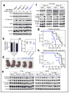
Figure 1
Figure 1. Characterization of RagAGTP/GTP mice
(A) Mouse embryo fibroblasts (MEFs) of RagA+/+, RagAGTP/+ and RagAGTP/GTP genotypes were deprived of amino acids for 1 h and re-stimulated for 10 min. Whole cell protein lysates were immunoblotted for indicated proteins. (B) Total RNA was extracted from RagA+/+ (n=3), RagAGTP/+ (n=3), and RagAGTP/GTP (n=2) MEFs and RagA mRNA expression determined by qPCR (mean ± SEM). (C) Weights of RagA+/+ (n=24), RagAGTP/+ (n=52), and RagAGTP/GTP (n=22) mice at birth (data are scatter dots, mean ± SD). (D) Representative photos of RagA+/+, RagAGTP/+, and RagAGTP/GTP neonates. Bar indicates 1 cm. (E) Early suppression of mTORC1 signaling after birth was determined by immunoblotting of protein extracts from liver and heart of RagA+/+ (+/+), RagAGTP/+ (G/+), and RagAGTP/GTP (G/G) neonates immediately after C-section (0 h) or after 1 h of fasting (1 h fast). (F) Liver and heart extracts from 10 h-fasted RagA+/+, RagAGTP/+, and RagAGTP/GTP neonates were analyzed by immunoblotting for the indicated proteins. (G) Survival curve of fasted neonates. Neonates obtained by C-section and resuscitated were fasted and their survival monitored (+/+: n=13; G/+: n=26; G/G: n=10). (H) Survival curve of fasted neonates treated with rapamycin. Neonates obtained by C-section and resuscitated were fasted and their survival monitored (untreated: n=10, rapamycin: n=6). Numbers indicate the median survival for each curve. *: p<0.05; **: p<0.01; ***: p<0.005.
Figure 2
Figure 2. Profound glucose homeostasis defect in RagAGTP/GTP mice
(A) Glycaemia drop in RagA+/+, RagAGTP/+ and RagAGTP/GTP and recovery in fasted RagA+/+ and RagAGTP/+ but not in RagAGTP/GTP neonates (+/+: n=5, 18, 4, 5, 9, 8; G/+: n=10, 26, 10, 13, 26, 21; G/G: n=7, 20, 9, 10, 16, 11, for 0, 1, 2, 3, 6 and 10 h, respectively; mean ± SEM). (B) Rapamycin significantly increases glycaemia in 6 h- and 10 h-fasted RagAGTP/GTP (mean ± SEM). (C) Extension of survival by glucose injections in fasted RagAGTP/GTP neonates (untreated: n=10; glucose: n=5). (D) Normal expression of genes involved in glucose metabolism in neonatal liver (+/+: n=2, G/+: n=5; G/G: n=4, mean ± SEM). (E) Left: Representative electron microscopy images showing abundant glycogen content in RagA+/+ and RagAGTP/GTP hepatocytes before fasting (0 h, upper panels) and more pronounced glycogen depletion after 10 h of fasting (lower panels) in RagAGTP/GTP neonates. Right: Quantification of hepatic glycogen content (+/+: n=5, 3, 4, 4; G/+: n=11, 7, 14, 15; G/G: n=6, 4, 4, 6; for 0, 1, 3, 6 and 10 h, respectively; mean ± SEM). (F) Partial rescue of hepatic glycogen content in 10 h-fasted RagAGTP/GTP neonates treated with rapamycin (mean ± SEM). (G) Quantification of neonatal plasma levels of branched-chain (BCAA) and essential amino acids at birth (left), after 10 h fasting (middle), and after 10 h fasting with rapamycin treatment (right) (n for +/+, G/+ and G/G, respectively: n=4, 5 and 4 for 0 h; n=4, 4 and 3 for 10 h; n=2, 5 and 3 for rapamycin; mean ± SD). Values are expressed relative to RagA+/+ levels at each time point. (H) Extension of survival by injection of a combination of gluconeogenic amino acids in fasted RagAGTP/GTP neonates (untreated: n=10; amino acids: n=8). *: p<0.05; **: p<0.01; ns: p>0.05.
Figure 3
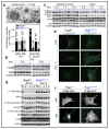
Figure 3. Impaired autophagy in RagAGTP/GTP neonates
(A) Top: Representative micrographs of autophagosomes and autophagolysosomes in hepatocytes from 1 h fasted RagA+/+ neonates. Typical autophagosome with a double limiting membrane (arrows); autophagosome and several autolysosomes (arrowheads). Bar indicates 5 μm. Bottom: Frequency of the two types of organelles (early: autophagosomes; late: autophagolysosomes) detected in cell profiles of hepatocytes and skeletal myocytes from RagA+/+ and RagAGTP/GTP neonates. (B) Protein extracts from livers of neonates at C-section (0 h) and 10 h-fasted (10 h) were immunblotted for autophagy markers LC3B and p62. (C) Protein extracts from skeletal muscle and heart from neonates at C-section (0 h), 1 h- and 2 h-fasted, were immunoblotted for indicated proteins. (D) Triggering of autophagy by amino acid withdrawal in MEFs. MEFs were deprived of amino acids for the indicated time points, whole cell protein extracts were obtained and mTORC1 activity and autophagic activity determined by immunoblotting. (E) Recombinant LC3B-GFP was expressed in RagA+/+ and RagAGTP/GTP MEFs and LC3B localization, in the presence and absence of amino acids, monitored by fluorescence microscopy. LC3B-GFP clustering, indicative of autophagy, was observed in amino acid-starved RagA+/+ but not RagAGTP/GTP MEFs. Bar indicates 10 μm. (F) Localization of recombinant TFEB-GFP was determined in MEFs as in (E). Nuclear (active) TFEB was observed in RagA+/+ MEFs upon amino acid withdrawal.
Figure 4
Figure 4. The Rag GTPases mediate inhibition of mTORC1 by glucose deprivation
(A) AMPKα1/α2 double knock-out (DKO) and wt MEFs were deprived of glucose for 1 h and re-stimulated for 10 min. Whole cell extracts were immunoblotted for the indicated proteins. (B) Immortalized MEFs of the indicated genotypes were deprived of growth factors, glucose, amino acids, or glucose and amino acids for 1 h and re-stimulated with glucose and/or amino acids for 10 min. Whole cell lysates were immunoblotted for the indicated proteins. (C) RagA+/+ and RagAGTP/GTP immortalized MEFs were deprived of glucose or amino acids and surviving cells quantified in triplicate after 48 h. Cell number is indicated relative to cell number at the start of the treatment; mean ± SD; ***: p<0.005. (D) mTOR localization as detected by immunofluorescence. In HEK-293T cells, glucose deprivation causes mTOR to localize to diffuse small puncta throughout the cytoplasm. Re-addition of glucose leads to mTOR shuttling to the lysosomal surface, co-localizing with the lysosomal protein Lamp2. HEK-293T-RagBGTP cells show mTOR localized at the lysosomal surface, regardless of glucose levels. Bar indicates 10 μm. (E) Glucose and amino acids affect the binding of the v-ATPase to the Ragulator complex. HEK-293T expressing FLAG-p14 were deprived of glucose or amino acids for 90 min and re-stimulated for 20 min. Protein extracts and immunoprecipitates were immunoblotted for the indicated proteins. (F) RagA+/+ and RagAGTP/GTP primary MEFs were cultured for 1 h in media with the glucose and amino acid concentrations measured in neonates at birth (0 h) or after 1 h fasting (1 h) and whole cells protein extracts were analyzed by immunoblotting. (G) Speculative model for constitutive RagA-induced neonatal lethality. Green and red boxes indicate active and inactive protein or process, respectively.

References

    1. Zoncu R, Efeyan A, Sabatini DM. mTOR: from growth signal integration to cancer, diabetes and ageing. Nat Rev Mol Cell Biol. 2010;12:21–35. doi: 10.1038/nrm3025. nrm3025 [pii] - DOI - PMC - PubMed
    1. Kim E, Goraksha-Hicks P, Li L, Neufeld TP, Guan KL. Regulation of TORC1 by Rag GTPases in nutrient response. Nat Cell Biol. 2008;10:935–945. doi: 10.1038/ncb1753. ncb1753 [pii] - DOI - PMC - PubMed
    1. Sancak Y, et al. The Rag GTPases bind raptor and mediate amino acid signaling to mTORC1. Science. 2008;320:1496–1501. doi: 10.1126/science.1157535. 1157535 [pii] - DOI - PMC - PubMed
    1. Sancak Y, et al. Ragulator-Rag complex targets mTORC1 to the lysosomal surface and is necessary for its activation by amino acids. Cell. 2010;141:290–303. doi: 10.1016/j.cell.2010.02.024. S0092-8674(10)00177-7 [pii] - DOI - PMC - PubMed
    1. Zoncu R, et al. mTORC1 senses lysosomal amino acids through an inside-out mechanism that requires the vacuolar H-ATPase. Science. 2011;334:678–683. doi: 10.1126/science.1207056. 334/6056/678 [pii] - DOI - PMC - PubMed

Publication types

MeSH terms