. 2013 Mar 15;97(4):666-75.
doi: 10.1093/cvr/cvs425. Epub 2012 Dec 21.

Hierarchical accumulation of RyR post-translational modifications drives disease progression in dystrophic cardiomyopathy

Affiliations

Hierarchical accumulation of RyR post-translational modifications drives disease progression in dystrophic cardiomyopathy

Sergii Kyrychenko et al. Cardiovasc Res. .

Abstract

Aims: Duchenne muscular dystrophy (DMD) is a muscle disease with serious cardiac complications. Changes in Ca(2+) homeostasis and oxidative stress were recently associated with cardiac deterioration, but the cellular pathophysiological mechanisms remain elusive. We investigated whether the activity of ryanodine receptor (RyR) Ca(2+) release channels is affected, whether changes in function are cause or consequence and which post-translational modifications drive disease progression.

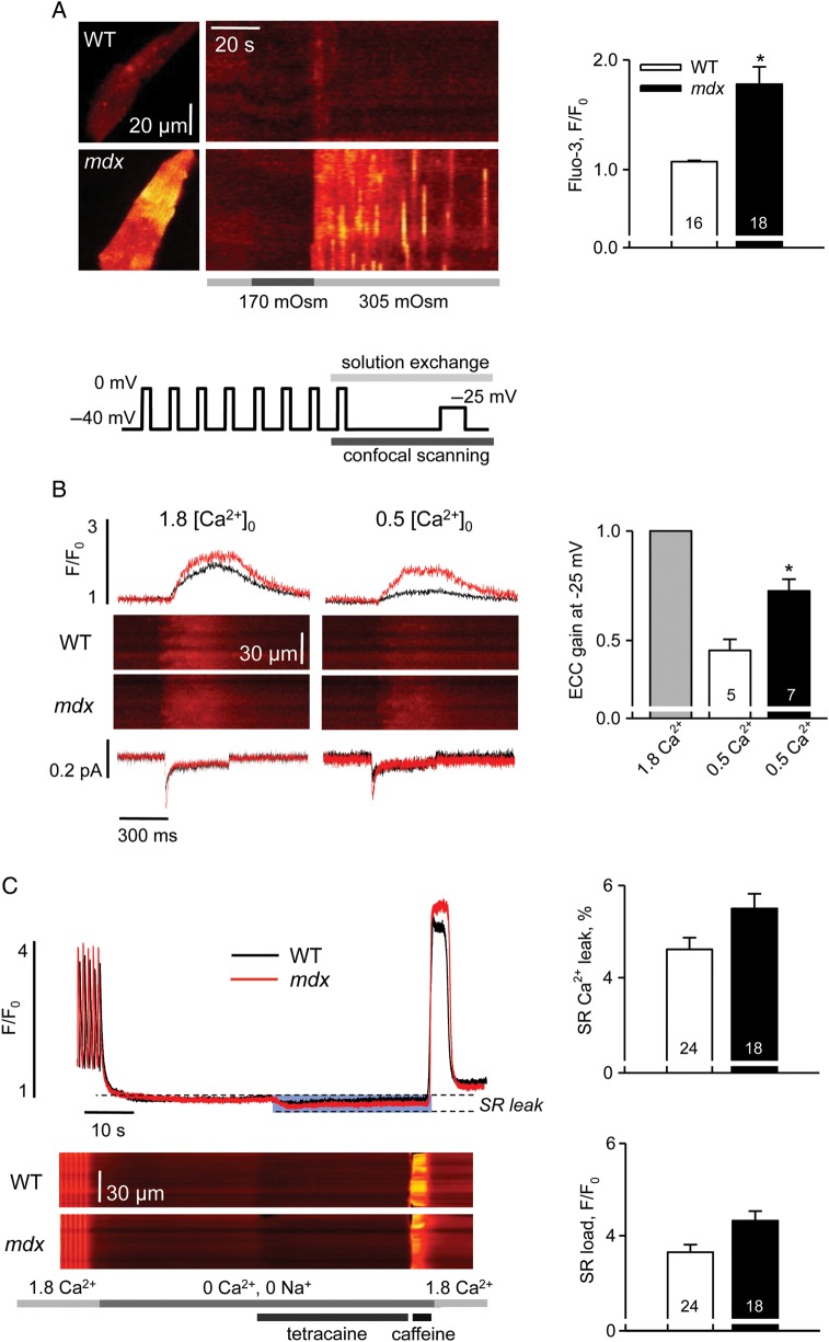
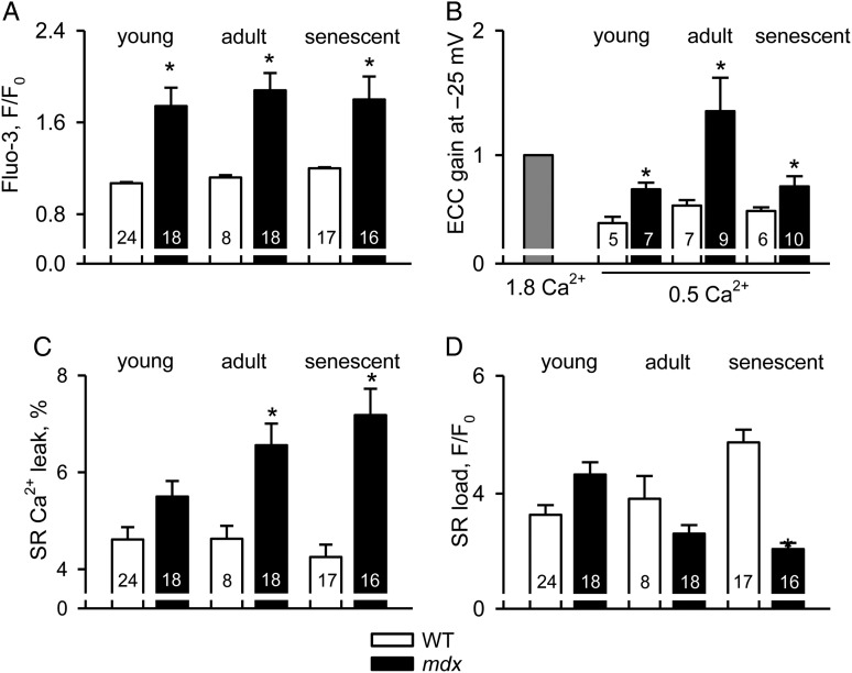
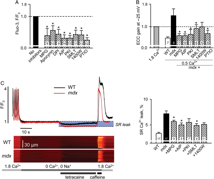
Methods and results: Electrophysiological, imaging, and biochemical techniques were used to study RyRs in cardiomyocytes from mdx mice, an animal model of DMD. Young mdx mice show no changes in cardiac performance, but do so after ∼8 months. Nevertheless, myocytes from mdx pups exhibited exaggerated Ca(2+) responses to mechanical stress and 'hypersensitive' excitation-contraction coupling, hallmarks of increased RyR Ca(2+) sensitivity. Both were normalized by antioxidants, inhibitors of NAD(P)H oxidase and CaMKII, but not by NO synthases and PKA antagonists. Sarcoplasmic reticulum Ca(2+) load and leak were unchanged in young mdx mice. However, by the age of 4-5 months and in senescence, leak was increased and load was reduced, indicating disease progression. By this age, all pharmacological interventions listed above normalized Ca(2+) signals and corrected changes in ECC, Ca(2+) load, and leak.

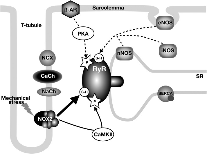
Conclusion: Our findings suggest that increased RyR Ca(2+) sensitivity precedes and presumably drives the progression of dystrophic cardiomyopathy, with oxidative stress initiating its development. RyR oxidation followed by phosphorylation, first by CaMKII and later by PKA, synergistically contributes to cardiac deterioration.


Figures

Figure 1
Figure 1
Intracellular calcium homeostasis in cardiomyocytes from 1-month-old mice. (A) Intracellular Ca2+ responses to mild hypo-osmotic shock in WT and mdx cells. Left panels are XY images of cardiac myocytes after returning to isotonic solution and line-scan representations of series of images acquired from the cells on the left upon application of an osmotic challenge. Averaged fluorescence was determined within each cell and converted to a two-dimensional X,t image (as in Martins et al.). Bars under the line-scans depict the protocol of extracellular solution changes. Right panel represents pooled data of mean values of normalized fluorescence during 60 s after the osmotic shock. The averaged response to the osmotic shock was extremely small in WT cells compared with mdx. (B) Left panels show representative traces of Ca2+ currents, line-scan images of Ca2+-related fluorescence and normalized cytosolic transients elicited by a 400 ms test pulse to −25 mV in WT and mdx cells superfused with either 1.8 or 0.5 mM Ca2+. Line plot on the top represents the voltage protocol used for the experiments. Right panel shows the statistical comparison of EC-coupling gain in 0.5 mM Ca2+ in WT and mdx cells. For each group, data were normalized to the value of the gain obtained in 1.8 mM Ca2+. SR Ca2+ release was much more resistant to the reduction in ICa trigger in mdx myocytes. (C) Left panels illustrate intracellular Ca2+ signals during the protocol designed to estimate SR Ca2+ leak (as in Shannon et al.): line-scan images of fluo-3 fluorescence and normalized cytosolic transients. Bars on the bottom depict the protocol of extracellular solution changes. Averaged values of estimated SR Ca2+ leak and SR Ca2+ load in WT and mdx cells are shown at the right. The SR Ca2+ leak was determined as a reduction in the resting fluo-3 fluorescence following tetracaine application, expressed as a per cent of SR Ca2+ content estimated from the amplitude of the caffeine-induced SR Ca2+ transient. There was no significant difference in the values obtained in WT and mdx cells. See Supplementary material online, Table S1 for details.
Figure 2
Figure 2
Post-translational modifications of RyRs in cells from 1-month-old mice. (A) Representative images of DCF fluorescence in WT and mdx myocytes under resting conditions and after application of 10 mM H2O2. Graph at the bottom left illustrates changes in the averaged DCF signals. Bar graphs compare the rate of DCF oxidation (slope) at rest and normalized increases in DCF signals after application of H2O2 in WT and mdx cells. The values were 0.027 ± 0.004 and 0.039 ± 0.003 for the slope and 18.93 ± 1.81 and 15.14 ± 1.33 for F(max)/F0 in WT and mdx cells, respectively. Both groups of measurements indicate higher ROS production in mdx cells. (B) Representative Coomassie-stained gel and mBB fluorescence intensity blot in samples from WT and mdx hearts. Bar plot shows averaged data on free thiol content in RyRs from WT myocytes normalized to the corresponding value in mdx cells. The level of free thiols is significantly greater in WT hearts. (C) Immunoblots and summary of oxidized CaMKII. Band intensities recorded from mdx samples were normalized per GAPDH signals and expressed as a percentage of increase compared with values obtained in WT samples. The level of oxidized CaMKII are almost two-fold higher in mdx hearts. (D) Immunoblot and summary of phosphorylated RyR. Whereas normalized intensity of CaMKII-dependent immunoreactivity increased ∼3-fold in dystrophic tissue, the value did not change significantly for the PKA site. Numbers of samples studied are indicated on the bars. (E) Reduction in oxidative stress and CaMKII inhibition suppress exaggerated intracellular Ca2+ responses to osmotic shock (left panel) and prevent hypersensitivity of EC-coupling (right panel). In the experiments with osmotic shock, individual intracellular Ca2+ responses were averaged within each experimental group (e.g. each pharmacological intervention applied) and normalized to the averaged response under control conditions (no drug applied). In the EC-coupling gain experiments, averaged data within each experimental group were also normalized to the values obtained in control (no drug applied) conditions. Details are in Supplementary material online, Table S1.
Figure 3
Figure 3
Gradual deterioration of intracellular Ca2+ homeostasis during the development of cardiac dystrophy. Intracellular Ca2+ responses to osmotic shock (A), gain of EC-coupling (B), intracellular Ca2+ leak (C), and SR Ca2+ content (D) in WT and mdx cardiomyocytes isolated from 1-, 3–4-, and 12–15-month-old mice. Note the gradually increased SR Ca2+ leak and decreased SR Ca2+ load in mdx cardiomyocytes from older mice. The corresponding values are listed in Supplementary material online, Table S2.
Figure 4
Figure 4
Reduction in oxidative/nitrosative stress as well as CaMKII and PKA inhibition ameliorates excessive intracellular Ca2+ responses to osmotic shock and hypersensitivity of EC-coupling in mdx myocytes isolated from 3- to 4-month-old mice (A and B) and reduces SR Ca2+ leak in cells from mature 12- 15-month-old mdx mice (C). In the experiments with osmotic shock (A), individual intracellular Ca2+ responses were determined as mean values of fluorescence recorded within the cell during 60 s after the shock, then averaged within each experimental group (e.g. each pharmacological intervention applied) and normalized to the averaged response under control conditions (no drug applied). In the EC-coupling gain experiments (B), averaged data within each experimental group were normalized to the values obtained in control (no drug applied) conditions. (C) Typical intracellular Ca2+ signals measured during the ‘leak’ protocol and averaged values of estimated SR Ca2+ leak under control conditions in WT and mdx cells and in mdx myocytes pretreated with various pharmacological agents (dashed bars). All values are listed in Supplementary material online, Table S1.
Figure 5
Figure 5
Signalling pathways involved in regulation of RyR activity in dystrophic cardiomyocytes. The diagram shows the main molecules involved in Ca2+ signalling and EC-coupling. Solid lines depict primary (oxidation and CaMKII-phosphorylation) and dashed lines depict secondary (nitrosation and PKA phosphorylation) mechanisms resulting in hypersensitivity of RyRs. CaCh and NaCh refer to L-type Ca2+ channel and voltage-gated Na+ channel, respectively.

References

    1. Hein S, Kostin S, Heling A, Maeno Y, Schaper J. The role of the cytoskeleton in heart failure. Cardiovasc Res. 2000;45:273–278. - PubMed
    1. Durbeej M, Campbell KP. Muscular dystrophies involving the dystrophin–glycoprotein complex: an overview of current mouse models. Curr Opin Genet Dev. 2002;12:349–361. - PubMed
    1. Finsterer J, Stöllberger C. The heart in human dystrophinopathies. Cardiology. 2003;99:1–19. - PubMed
    1. Quinlan JG, Hahn HS, Wong BL, Lorenz JN, Wenisch AS, Levin LS. Evolution of the mdx mouse cardiomyopathy: physiological and morphological findings. Neuromuscul Disord. 2004;14:491–496. - PubMed
    1. Stuckey DJ, Carr CA, Camelliti P, Tyler DJ, Davies KE, Clarke K. In vivo MRI characterization of progressive cardiac dysfunction in the mdx mouse model of muscular dystrophy. PLoS One. 2012;7:e28569. - PMC - PubMed

Publication types

MeSH terms

Substances