. 2013 Feb;16(2):174-82.
doi: 10.1038/nn.3289. Epub 2012 Dec 23.

A subpopulation of nociceptors specifically linked to itch

Affiliations

A subpopulation of nociceptors specifically linked to itch

Liang Han et al. Nat Neurosci. 2013 Feb.

Abstract

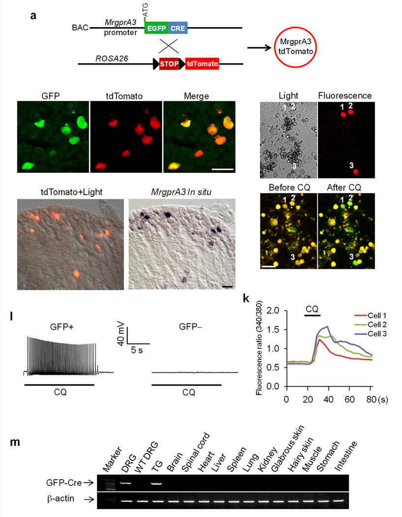
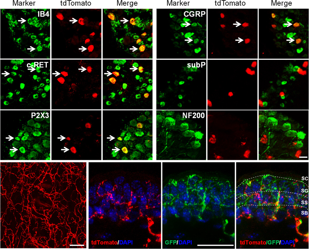
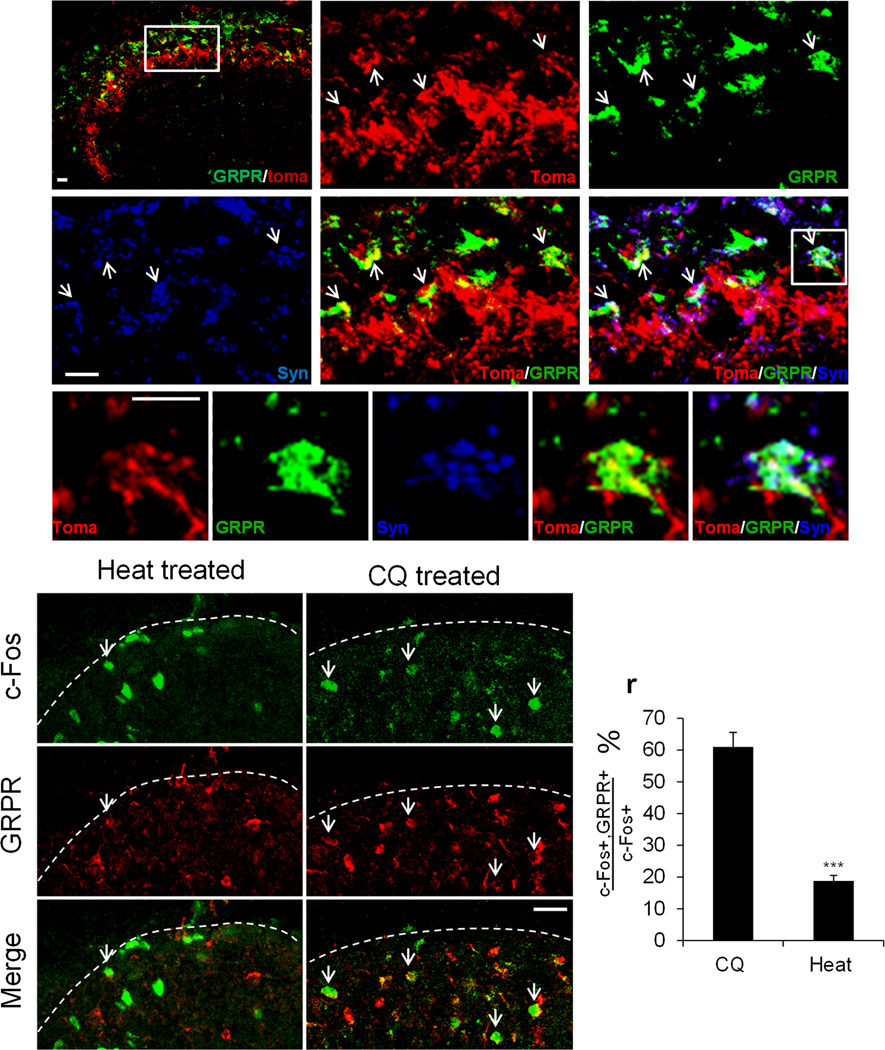
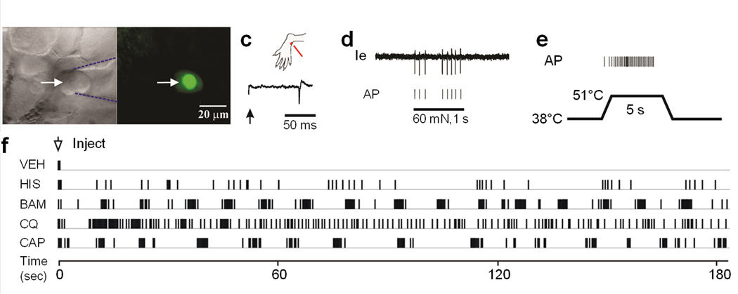
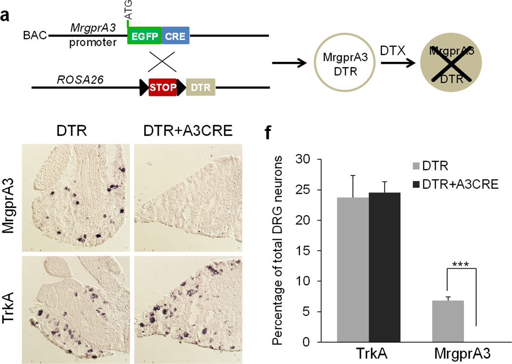
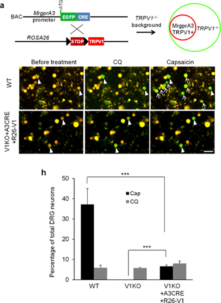
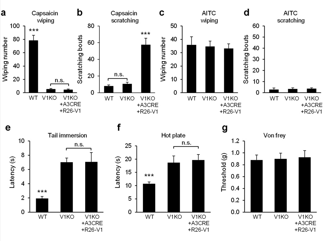
Itch-specific neurons have been sought for decades. The existence of such neurons has been doubted recently as a result of the observation that itch-mediating neurons also respond to painful stimuli. We genetically labeled and manipulated MrgprA3(+) neurons in the dorsal root ganglion (DRG) and found that they exclusively innervated the epidermis of the skin and responded to multiple pruritogens. Ablation of MrgprA3(+) neurons led to substantial reductions in scratching evoked by multiple pruritogens and occurring spontaneously under chronic itch conditions, whereas pain sensitivity remained intact. Notably, mice in which TRPV1 was exclusively expressed in MrgprA3(+) neurons exhibited itch, but not pain, behavior in response to capsaicin. Although MrgprA3(+) neurons were sensitive to noxious heat, activation of TRPV1 in these neurons by noxious heat did not alter pain behavior. These data suggest that MrgprA3 defines a specific subpopulation of DRG neurons mediating itch. Our study opens new avenues for studying itch and developing anti-pruritic therapies.


Figures

Figure 1
Figure 1. Generation of the MrgprA3GFP-Cre transgenic mouse line
(a) Diagram showing the mating strategy. (b–d) DRG sections from an MrgprA3GFP-Cre; ROSA26tdTomato mouse stained with anti-GFP antibody. TdTomato fluorescence was visualized directly without staining. (e) Merged image of tdTomato fluorescence and bright field view of a DRG section from a MrgprA3GFP-Cre; ROSA26tdTomato mouse. (f) In situ hybridization with an MrgprA3 probe on the same section shown in (e). (g–j) Representative light view (g), fluorescent view (h), and Fura-2 ratiometric images of tdTomato+ DRG neurons (i,j). The color of the neurons switching from yellow to green indicates the increase of the intracellular calcium concentration. (k) Representative traces evoked by chloroquine (CQ; 1 mM) in the calcium imaging assay from the three tdTomato+ neurons labeled in (j). (l) Representative traces of APs induced by chloroquine (1 mM) in neurons from MrgprA3GFP-Cre mice. chloroquine can induce APs in GFP-Cre+ DRG neurons (10 of 11). In contrast, GFP-Cre DRG neurons (12) do not show any response to chloroquine. (m) RT-PCR analysis of GFP-Cre expression in various tissues from MrgprA3GFP-Cre mice. GFP-Cre was only detected in the DRG and trigeminal ganglia. Scale bar represents 50 μm.
Figure 2
Figure 2. Characterization of MrgprA3+ neurons
(a–r) L4-L6 DRG sections from MrgprA3GFP-Cre; ROSA26tdTomato mice stained with the indicated markers. Arrows mark representative double-labeled neurons. (s) Whole-mount imaging of the dorsal thoracic skin from MrgprA3GFP-Cre; ROSA26tdTomato mice showing the distribution of MrgprA3+ nerve fibers. (t–v) Section of the dorsal thoracic hairy skin from MrgprA3GFP-Cre; ROSA26tdTomato; MrgprDGFP/+ mice showing MrgprA3+ and MrgprD+ free nerve endings in the superficial epidermis. The section was counterstained with DAPI to label nuclei. Scale bar represents 30 μm. SC, stratum corneum; SG, stratum granulosum; SB, stratum basalis; SS, stratum spinosum. The epidermal subdivisions were identified by keratinocyte nuclear morphology and packing density.
Figure 3
Figure 3. MrgprA3+ DRG neurons form synaptic connections with GRPR+ neurons in the dorsal spinal cord
(a–k) spinal cord cross-sections from the thoracic region of adult MrgprA3GFP-Cre; ROSA26tdTomato mice labeled with GRPR (green), tdTomato reporter (red) and presynaptic marker synapsin 1 (blue).The boxed area in a is shown at greater magnification in b–f. The representative neuron in boxed region in f is shown at greater magnification in g–k. Scale bars, 10 µm. Arrows point to representative synaptic connections between tdTomato+ nerve fiber and GRPR+ neurons. (l–q) Double labeling of c-Fos-GFP (green) and GRPR (red) on the L3-L5 spinal cord cross-sections from c-FosGFP mice treated with chloroquine (8 mM) or hot water (50°C). Arrows indicate double labeled cells. Dashed lines define the boundary of spinal cord sections. (r) The percentage of c-Fos+ neurons that coexpress GRPR in the ipsilateral superficial spinal cords. Majority of c-Fos+ neurons induced by chloroquine are GRPR+ dorsal horn neurons whereas majority of heat-induced c-Fos+ neurons do not express GRPR (n=3, p=0.004). Error bars represent SEM.
Figure 4
Figure 4. MrgprA3+ neurons have polymodal nociceptors with C-fibers and respond to multiple pruritogens
(a–f) In-vivo electrophysiological recording of MrgprA3+ neurons. (a) Bright field image of a neuronal recording (arrow) with an extracellular electrode (outlined with dashed blue lines). (b) Fluorescent microscopy revealed the expression of GFP (nuclear GFP fluorescence in the MrgprA3+ neuron. (c) Location of the cutaneous receptive field (RF, red dot) of this neuron on the hairy skin of the hindpaw and conduction velocity (lower trace, 0.49 m/s for the MrgprA3+ neuron), obtained with electrical stimulation (arrow) of the RF, are shown. (d) Responses of the neuron to a 60 mN force via a 200 μm diameter probe applied to its RF (1 sec) with original extracellular recording trace (Ie) and each action potential (AP) indicated by the vertical tic mark below. (e) Response to heat stimulation (38 to 51°C, 5 sec) indicates that this is a CMH neuron. (f) Responses of the CMH neuron to the intradermal injection for the MrgprA3 neuron of vehicle (VEH), followed by histamine (HIS, 5.4 mM), BAM8–22 (BAM, 0.2 mM), chloroquine (CQ, 1mM), and capsaicin (CAP, 3.3 mM).
Figure 5
Figure 5. The ablation of MrgprA3+ neurons
(a) Diagram showing the mating strategy. (b–e) In situ hybridizations of DRG sections from DTX-treated MrgprA3GFP-Cre; ROSA26DTR and ROSA26DTR littermates using MrgprA3 (b, c) and TrkA (d, e) cDNA probes demonstrate that MrgprA3+ neurons were specifically ablated in MrgprA3GFP-Cre; ROSA26DTRmice. (f) Quantitation of the in situ hybridization results (n = 3 mice per genotype, MrgprA3, p = 0.004, TrkA, p = 0.80). ***p < 0.005; two-tailed unpaired Student’s t-test.
Figure 6
Figure 6. Specific activation of MrgprA3+ neurons
(a) Diagram showing the mating strategy. MrgprA3GFP-Cre; ROSA26TRPV1 mice were generated in TRPV1 knockout background to exclusively express TRPV1 in MrgprA3+ neurons. (b–g) Fura-2 ratiometric images of cultured DRG neurons from WT (b–d) and TRPV1−/−; MrgprA3GFP-Cre; ROSA26TRPV1 mice. The color of the neurons switching from yellow to green indicates the increase of the intracellular calcium concentration (b–g). In WT DRG neurons, capsaicin (1µM) activated a much bigger population than chloroquine (1mM). However, the percentage of TRPV1−/−; MrgprA3GFP-Cre; ROSA26TRPV1 DRG neurons responding to capsaicin and chloroquine are similar and every capsaicin-responding neuron responded to chloroquine. Arrowheads point to the neurons responded to both chloroquine and capsaicin. Arrows point to the neurons only responded to capsaicin. (h) The percentage of total DRG neurons from WT, TRPV1−/− mice, and TRPV1−/−; MrgprA3GFP-Cre; ROSA26TRPV1 mice responded to chloroquine (1mM) and capsaicin (1µM). ***p < 0.005; two-tailed unpaired Student’s t-test.
Figure 7
Figure 7. Specific activation of MrgprA3+ neurons evokes robust scratching and little or no pain response
(a) Cheek injection of capsaicin (3.3 mM induced robust, site-directed wiping with the forepaw in WT mice but not in TRPV1−/− mice or TRPV1−/−; MrgprA3GFP-Cre; ROSA26TRPV1 mice (n=13 vs. 13 vs. 10, WT vs. V1KO, p=1.8E-09, V1KO vs. V1KO+A3CRE+R26-V1, p=0.59). (b) Cheek injection of capsaicin (3.3 mM) induced robust site-directed scratching with the hind paw in TRPV1−/−; MrgprA3GFP-Cre; ROSA26TRPV1 mice but not in WT mice or TRPV1−/− mice (WT vs. V1KO vs. V1KO+A3CRE+R26-V1, n=13 vs. 13 vs.10, WT vs. V1KO, p=0.28, V1KO vs. V1KO+A3CRE+R26-V1, p=7.2E-07). (c–d) Cheek injection of allyl isothiocyanate (AITC, 50mM) induced robust wiping (c), not scratching (d) (WT vs. V1KO vs. V1KO+A3CRE+R26-V1, n=8 vs. 8 vs.7). (e) Response latencies of WT, TRPV1−/− and TRPV1−/−; MrgprA3GFP-Cre; ROSA26TRPV1 mice in the tail immersion test (50°C) (WT vs. V1KO vs. V1KO+A3CRE+R26-V1, n=10 vs. 11 vs.8, WT vs. V1KO, p=2.3E-07, V1KO vs. V1KO+A3CRE+R26-V1, p=0.97). (f) Response latencies in the hot plate tests (55°C) (WT vs. V1KO vs. V1KO+A3CRE+R26-V1, n=11 vs. 8 vs. 7, WT vs. V1KO, p=0.0017, V1KO vs. V1KO+A3CRE+R26-V1, p=0.75). (g) Response threshold in the von Frey test (WT vs. V1KO vs. V1KO+A3CRE+R26-V1, n=10 vs. 10 vs. 8, WT vs. V1KO, p=0.87, V1KO vs. V1KO+A3CRE+R26-V1, p=0.86). ***p < 0.005; two-tailed unpaired Student’s t-test. n.s., not significant.

Comment in

  • Scratching an itch.
    Namer B, Reeh P. Namer B, et al. Nat Neurosci. 2013 Feb;16(2):117-8. doi: 10.1038/nn.3316. Nat Neurosci. 2013. PMID: 23354383 No abstract available.

References

    1. Ikoma A, Steinhoff M, Stander S, Yosipovitch G, Schmelz M. The neurobiology of itch. Nat Rev Neurosci. 2006;7:535–547. - PubMed
    1. Basbaum AI, Bautista DM, Scherrer G, Julius D. Cellular and molecular mechanisms of pain. Cell. 2009;139:267–284. - PMC - PubMed
    1. Schmelz M, Schmidt R, Bickel A, Handwerker HO, Torebjork HE. Specific C-receptors for itch in human skin. J Neurosci. 1997;17:8003–8008. - PMC - PubMed
    1. Schmelz M, et al. Chemical response pattern of different classes of C-nociceptors to pruritogens and algogens. J Neurophysiol. 2003;89:2441–2448. - PubMed
    1. Johanek LM, et al. A role for polymodal C-fiber afferents in nonhistaminergic itch. J Neurosci. 2008;28:7659–7669. - PMC - PubMed

Publication types

MeSH terms

Substances