. 2013 Jan;19(1):74-82.
doi: 10.1038/nm.3040. Epub 2012 Dec 23.

An endothelial apelin-FGF link mediated by miR-424 and miR-503 is disrupted in pulmonary arterial hypertension

Affiliations

An endothelial apelin-FGF link mediated by miR-424 and miR-503 is disrupted in pulmonary arterial hypertension

Jongmin Kim et al. Nat Med. 2013 Jan.

Abstract

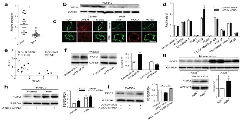
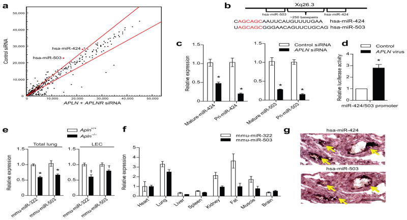
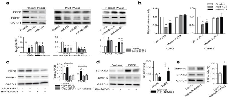
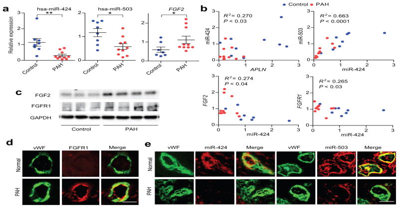
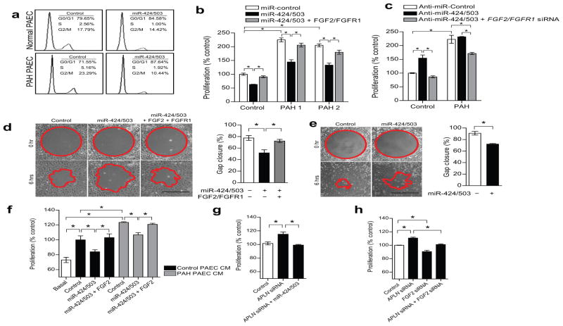
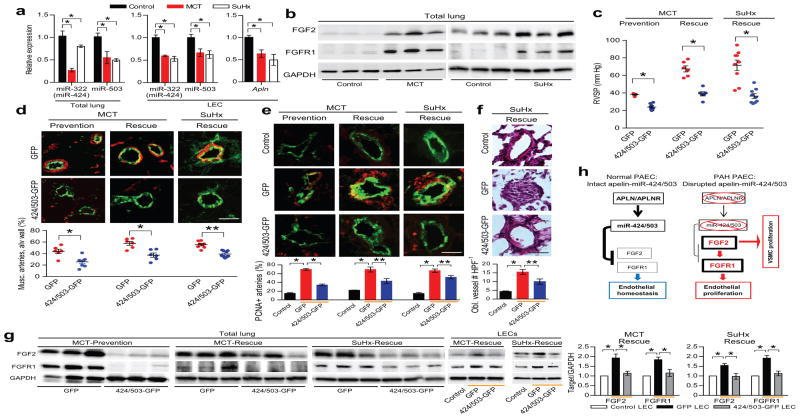
Pulmonary arterial hypertension (PAH) is characterized by vascular remodeling associated with obliteration of pulmonary arterioles and formation of plexiform lesions composed of hyperproliferative endothelial and vascular smooth-muscle cells. Here we describe a microRNA (miRNA)-dependent association between apelin (APLN) and fibroblast growth factor 2 (FGF2) signaling in pulmonary artery endothelial cells (PAECs). APLN deficiency in these cells led to increased expression of FGF2 and its receptor FGFR1 as a consequence of decreased expression of miR-424 and miR-503, which directly target FGF2 and FGFR1. miR-424 and miR-503 were downregulated in PAH, exerted antiproliferative effects in PAECs and inhibited the capacity of PAEC-conditioned medium to induce the proliferation of pulmonary artery smooth-muscle cells. Reconstitution of miR-424 and miR-503 in vivo ameliorated pulmonary hypertension in experimental models. These studies identify an APLN-dependent miRNA-FGF signaling axis needed for the maintenance of pulmonary vascular homeostasis.


Conflict of interest statement

Competing financial interests

The authors declare no competing financial interests.

Figures

Figure 1
Figure 1. APLN reciprocally regulates FGF2 expression in PAECs in a miRNA dependent manner
(a) APLN mRNA expression in PAECs of controls and PAH subjects. *P < 0.005 vs. controls. (b) APLN protein expression in PAECs of controls and PAH subjects. (c) Immunofluorescence staining showing APLN expression and PCNA staining in the lung endothelium of a control and a PAH subject. Von Willebrand Factor (vWF) staining is shown in green, APLN and PCNA staining are shown in red. Scale bar = 50 μm. (d) Cytokine array showing relative changes in cytokine expression in response to APLN knockdown in PAECs. *P < 0.01 vs. control siRNA. (e) Correlation between APLN and FGF2 mRNA levels in PAECs from normal controls and PAH subjects. (f) Expression of FGF2 protein in response to either APLN knockdown or lentiviral APLN overexpression in PAECs. *P < 0.01. (g) Expression of FGF2 protein in total lung homogenates and isolated LECs from wildtype and Apln null mice. *P < 0.05. (h) FGF2 expression in response to knockdown of AGO2 in normal and PAH PAECs. *P < 0.01. (i) FGF2 expression in PAECs in response to APLN overexpression and AGO2 knockdown. *P < 0.01.
Figure 2
Figure 2. APLN-FGF2 reciprocal axis is mediated by APLN regulated expression of miR-424 and miR-503
(a) MiRNA microarray analysis of PAECs subjected to APLN and APLNR knockdown. MiR-424 and miR-503 are depicted in red. Red lines demarcate a 1.2 fold change from baseline expression. (b) Chromosomal location of miR-424 and miR-503 and the sequences of the mature miRNAs. The homology in the seed sequences is depicted in red. (c) Quantitative PCR showing expression of the mature and pri-forms of miR-424 and miR-503 in response to APLN knockdown in PAECs. *P < 0.01 vs. control siRNA. (d) Relative luciferase activity of PAECs transfected with the putative miR-424 and miR-503 promoter based luciferase reporter construct in response to APLN overexpression. *P < 0.001. (e) Expression of mmu-miR-322 (mouse homolog of hsa-miR-424) and mmu-miR-503 expression in total lungs and LECs of Apln deficient mice. *P < 0.001 and †P < 0.01. (f) Quantitative PCR of mmu-miR-322 and mmu-miR-503 of mouse tissues. (g) In situ hybridization of human lungs for miR-424 and miR-503. Yellow arrows depict positive staining cells. Scale bar = 100 μm.
Figure 3
Figure 3. MiR-424 and miR-503 regulate FGF2 and FGFR1 expression and signaling
(a) FGF2 and FGFR1 protein expression in response to either overexpression of miR-424 and miR-503 in normal and PAH PAECs, and inhibition of miR-424 or miR-503 in normal PAECs. *P < 0.01 and †P < 0.05. (b) Targeting of FGF2 and FGFR1 3′ UTR by miR-424 and miR-503. Both wildtype (WT) and mutagenized 3′ UTR constructs are shown. *P < 0.01 vs. control. (c) FGF2 and FGFR1 expression in response to knockdown of APLN in PAECs with concurrent overexpression of miR-424 and miR-503. *P < 0.01. (d) ERK1/2 phosphorylation in response to overexpression of miR-424 and miR-503 in PAECs, both at baseline and with FGF2 stimulation. *P < 0.05 and †P < 0.01. (e) ERK1/2 phosphorylation with inhibition of miR-424 and miR-503 with anti-miRs in PAECs. *P < 0.01.
Figure 4
Figure 4. Downregulation of miR-424 and miR-503 in PAH is associated with increased FGF2 and FGFR1 expression
(a) Expression of miR-424, miR-503, and FGF2 mRNA in PAECs of controls and PAH subjects. *P < 0.01, **P < 0.05. (b) Correlation plots for expression levels of APLN mRNA and miR-424, miR-424 and miR-503, FGF2 mRNA and miR-424, and FGFR1 mRNA and miR-424 in PAECs from control and PAH PAECs. (c) Expression of FGF2 and FGFR1 proteins in control and PAH PAECs. (d) FGFR1 expression (shown in red) in control and PAH lung microvascular endothelium with costaining for vWF (shown in green). (e) Expression of miR-424 and miR-503 by in situ hybridization (shown in red) in the pulmonary endothelium (vWF staining in green) in the microvasculature of control and PAH lungs. Scale bar = 50 μm.
Figure 5
Figure 5. Endothelial miR-424 and miR-503 regulate PAEC proliferation and migration and induce paracrine inhibition of PASMC proliferation
(a) Cell cycle analysis of PAECs with miR-424 and miR-503 overexpression in normal and PAH PAECs. (b) Proliferation of normal and PAH PAECs with overexpression of miR-424 and miR-503 or with concurrent stimulation with exogenous FGF2 and transfection of FGFR1 expression construct. *P < 0.01. (c) Proliferation of normal PAECs and PAH PAECs in response to inhibition of miR-424 and miR-503 by anti-miR transfection or with concurrent knockdown of FGF2 and FGFR1. *P < 0.01. (d) Cell migration response to miR-424 and miR-503 overexpression or with concurrent FGF2 stimulation and FGFR1 trasfection in normal PAECs. *P < 0.05 vs. controls. Scale bar = 500 μm. (e) Cell migration response to overexpression of miR-424 and miR-503 in PAH PAECs. *P < 0.05. Scale bar = 500 μm. (f) PASMC proliferation in response to conditioned media (CM) from PAECs of a normal donor and a PAH subject (Control vs. Basal), CM from respective PAECs transfected with miR-424 and miR-503 mimics, and co-transfected with FGF2 expression construct. *P < 0.01. (g) PASMC proliferation in response to CM from PAECs subjected to either APLN knockdown alone or in conjunction with miR-424 and miR-503 overexpression. *P < 0.05. (h) Proliferation of PASMCs in response to CM from PAECs subjected to APLN knockdown, FGF2 knockdown, or concurrent knockdown. *P < 0.05.
Figure 6
Figure 6. MiR-424 and miR-503 can ameliorate the experimental pulmonary hypertension models in rodents
(a) Expression levels of rno-miR-322 (hsa-miR-424 equivalent in rat) and miR-503 in rat lungs and isolated LECs 3 weeks after MCT injection and 4 weeks after SuHx. Expression levels of Apln mRNA in the rat LECs from two pulmonary hypertension models. *P < 0.01. (b) Expression of FGF2 and FGFR1 in the MCT and SuHx induced pulmonary hypertension rat lungs. (c) Right ventricular systolic pressure (RVSP) measurements in rats receiving intranasal delivery of lentiviral miR-424 and miR-503 (424/503-GFP) compared to control lentivirus (GFP) in the MCT prevention and rescue models, and the SuHx rescue model. *P < 0.001 for each model. (d) Microvascular muscularization analysis of lungs from rats receiving either intranasal GFP or 424/503-GFP in the three models. Smooth muscle actin is shown in red, vWF is shown in green. *P < 0.01 and **P < 0.001. (e) Assessment of PCNA expression in the lungs of the three pulmonary hypertension models receiving either GFP control or 424/503-GFP. PCNA is shown in red, vWF is shown in green. Orange bars denote the experimental pulmonary hypertension groups. *P < 0.001 and **P < 0.01. (f) H&E staining of the lungs from rats subjected to SuHx pulmonary hypertension induction receiving either GFP or 424/503-GFP. The average number of obliterated vessels per microscopic field is shown. Orange bars denote the experimental pulmonary hypertension groups. *P < 0.001, **P < 0.02. (g) FGF2 and FGFR1 protein levels in the lung homogenates of rats in the three models with either GFP or 424/503-GFP. FGF2 and FGFR1 expression in the isolated LECs from the MCT-Rescue and SuHx-Rescue models with GFP or 424/503-GFP. Orange bars denote the experimental pulmonary hypertension groups. *P < 0.05. (h) Proposed mechanism of endothelial signal linking APLN, miR-424/miR-503, and FGF2-FGFR1 in normal and PAH pulmonary endothelium.

Comment in

References

    1. Tuder RM, Marecki JC, Richter A, Fijalkowska I, Flores S. Pathology of pulmonary hypertension. Clin Chest Med. 2007;28:23–42. vii. - PMC - PubMed
    1. Humbert M, et al. Cellular and molecular pathobiology of pulmonary arterial hypertension. J Am Coll Cardiol. 2004;43:13S–24S. - PubMed
    1. Hassoun PM, et al. Inflammation, growth factors, and pulmonary vascular remodeling. J Am Coll Cardiol. 2009;54:S10–19. - PubMed
    1. Schermuly RT, Ghofrani HA, Wilkins MR, Grimminger F. Mechanisms of disease: pulmonary arterial hypertension. Nature reviews Cardiology. 2011;8:443–455. - PMC - PubMed
    1. Izikki M, et al. Endothelial-derived FGF2 contributes to the progression of pulmonary hypertension in humans and rodents. J Clin Invest. 2009;119:512–523. - PMC - PubMed

Publication types

MeSH terms

Associated data