Review
. 2013 Mar;81(3):629-35.
doi: 10.1128/IAI.01035-12. Epub 2012 Dec 21.

Tetratricopeptide repeat motifs in the world of bacterial pathogens: role in virulence mechanisms

Affiliations
Review

Tetratricopeptide repeat motifs in the world of bacterial pathogens: role in virulence mechanisms

Lukas Cerveny et al. Infect Immun. 2013 Mar.

Abstract

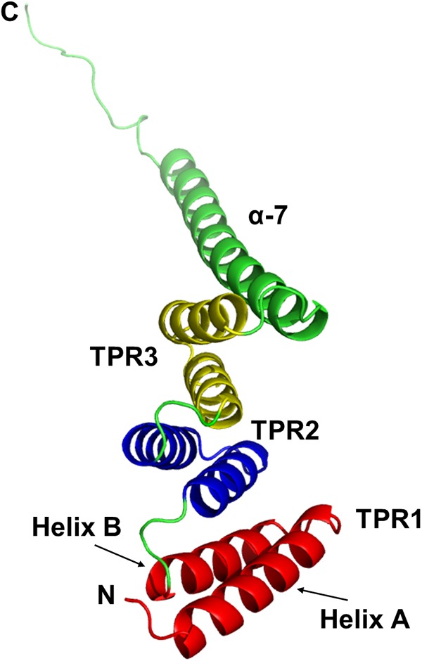
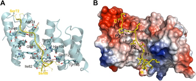
The tetratricopeptide repeat (TPR) structural motif is known to occur in a wide variety of proteins present in prokaryotic and eukaryotic organisms. The TPR motif represents an elegant module for the assembly of various multiprotein complexes, and thus, TPR-containing proteins often play roles in vital cell processes. As the TPR profile is well defined, the complete TPR protein repertoire of a bacterium with a known genomic sequence can be predicted. This provides a tremendous opportunity for investigators to identify new TPR-containing proteins and study them in detail. In the past decade, TPR-containing proteins of bacterial pathogens have been reported to be directly related to virulence-associated functions. In this minireview, we summarize the current knowledge of the TPR-containing proteins involved in virulence mechanisms of bacterial pathogens while highlighting the importance of TPR motifs for the proper functioning of class II chaperones of a type III secretion system in the pathogenesis of Yersinia, Pseudomonas, and Shigella.


Figures

Fig 1
Fig 1
Ribbon diagram of the TPR domain of PP5. PP5 was the first TPR-containing protein whose structure was determined by crystallography (5). We adopted this protein to illustrate the structure of the TPR-containing proteins. PP5 contains three tandem TPR motifs (TPR1 to TPR3), which are depicted in different colors. Each TPR motif is composed of a pair of antiparallel α-helices, termed helices A and B. The structures of all of the TPR motifs are virtually identical. The three TPR motifs are organized in a parallel arrangement, such that sequentially adjacent α-helices are antiparallel. The 35 residues C terminal to the three TPR motifs of PP5 are folded into an extended α-helix (α-7). This helix is packed against helix B of TPR3 in an arrangement similar to that of the helices within the TPR domain. The consequence of the regular repetition of such α-helices is the formation of a right-handed helical conformation that creates an amphipathic channel (5). The image shown was prepared with the PyMOL Molecular Graphics System, version 1.3, Schrödinger, LLC. (Adapted from reference by permission from Nature Publishing Group.)
Fig 2
Fig 2
TPR-containing proteins accommodate their cognate substrates in the concave cleft. For illustration, we adopted the interaction between the TTSS class II chaperone of Shigella, IpgC, and the chaperone binding motif of its cognate substrate, IpaB. IpgC binds the chaperone binding domain in an extended conformation that is stabilized by conserved residues present in the cleft. (A) Diagram representing IpgC and a yellow ribbon model of the chaperone binding domain of IpaB. The residues involved in the intermolecular interactions are shown. The H bonds (between the carbonyl of Ile-62, Pro-65, and Lys-68 in the IpaB peptide and the amide of Gln-112 and the hydroxyl of Tyr-47 and Tyr-40 in IpgC) and one salt bridge (Lys-68 in the IpaB peptide and Asp-71 in IpgC) are emphasized as dashed red lines. (B) Same view as in panel A showing a surface representation of IpgC in the cocrystal. The surface is colored according to the electrostatic potential calculated without water and ligand (blue, positive; red, negative). The chaperone binding domain of IpaB (yellow) lining the major groove of IpgC and the three pockets (P1 to P3) interacting with side chains of conserved residues Pro-65, Leu-67, and Pro-70 in the IpaB peptide via hydrophobic and van der Waals interactions, P1, P2, and P3, are indicated. The 65PELKAP70 residues of the IpaB peptide are labeled. (Adapted from reference with the permission of the publisher.)
Fig 3
Fig 3
Localization of TPR motifs identified by crystallography and/or predicted utilizing the web-based tool kits Pfam (30), SMART (31), and TPRpred (32). Protein names, bacterial origins, and UniProt database accession numbers are shown in columns 1 to 3, respectively. The motifs determined by the particular web-based tool kits are depicted in specific colors. Numbers refer to the first and last residues of the structural units.

References

    1. Hirano T, Kinoshita N, Morikawa K, Yanagida M. 1990. Snap helix with knob and hole: essential repeats in S. pombe nuclear protein nuc2+. Cell 60:319–328 - PubMed
    1. Sikorski RS, Boguski MS, Goebl M, Hieter P. 1990. A repeating amino acid motif in CDC23 defines a family of proteins and a new relationship among genes required for mitosis and RNA synthesis. Cell 60:307–317 - PubMed
    1. Lamb JR, Tugendreich S, Hieter P. 1995. Tetratrico peptide repeat interactions: to TPR or not to TPR? Trends Biochem. Sci. 20:257–259 - PubMed
    1. D'Andrea LD, Regan L. 2003. TPR proteins: the versatile helix. Trends Biochem. Sci. 28:655–662 - PubMed
    1. Das AK, Cohen PW, Barford D. 1998. The structure of the tetratricopeptide repeats of protein phosphatase 5: implications for TPR-mediated protein-protein interactions. EMBO J. 17:1192–1199 - PMC - PubMed

Publication types

MeSH terms

Substances

LinkOut - more resources