. 2013 Feb;19(2):129-40.
doi: 10.1261/rna.034777.112. Epub 2012 Dec 21.

Neurodegenerative diseases: quantitative predictions of protein-RNA interactions

Affiliations

Neurodegenerative diseases: quantitative predictions of protein-RNA interactions

Davide Cirillo et al. RNA. 2013 Feb.

Abstract

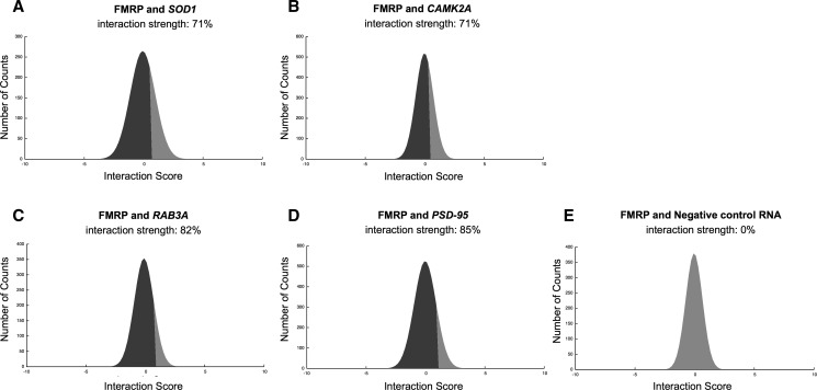
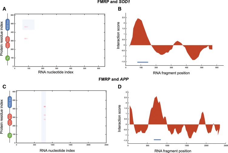
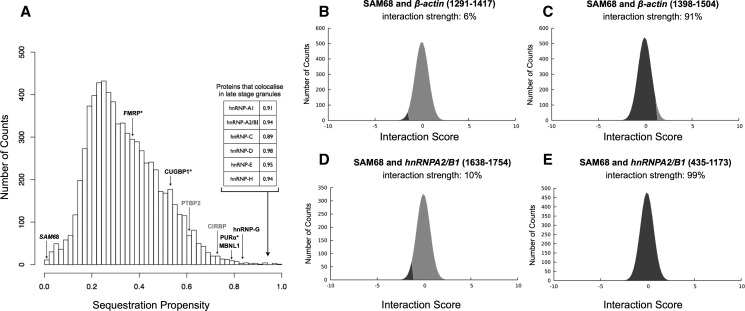
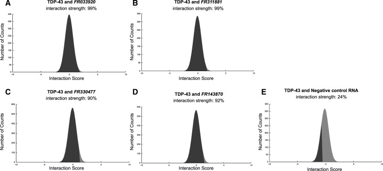
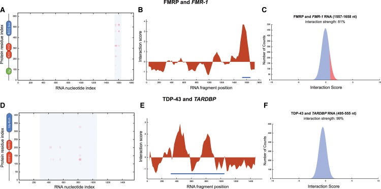
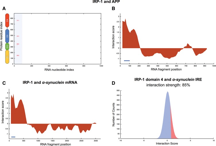
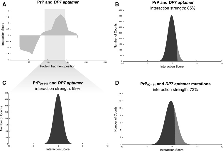
Increasing evidence indicates that RNA plays an active role in a number of neurodegenerative diseases. We recently introduced a theoretical framework, catRAPID, to predict the binding ability of protein and RNA molecules. Here, we use catRAPID to investigate ribonucleoprotein interactions linked to inherited intellectual disability, amyotrophic lateral sclerosis, Creutzfeuld-Jakob, Alzheimer's, and Parkinson's diseases. We specifically focus on (1) RNA interactions with fragile X mental retardation protein FMRP; (2) protein sequestration caused by CGG repeats; (3) noncoding transcripts regulated by TAR DNA-binding protein 43 TDP-43; (4) autogenous regulation of TDP-43 and FMRP; (5) iron-mediated expression of amyloid precursor protein APP and α-synuclein; (6) interactions between prions and RNA aptamers. Our results are in striking agreement with experimental evidence and provide new insights in processes associated with neuronal function and misfunction.


Figures

FIGURE 1.
FIGURE 1.
FMRP associations with coding transcripts. Prediction of FMRP association with (A) SOD1, (B) CAMK2A, (C) RAB3A, (D) PSD-95 mRNAs, and (E) negative control RNA. The interaction strength is the propensity to bind with respect to a reference set of protein–RNA associations (black area in the score distribution; Supplemental Table 1).
FIGURE 2.
FIGURE 2.
FMRP interactions with SOD1 and APP. Interaction maps of FMRP binding to (A) SOD1 and (C) APP (FMRP secondary structure elements are displayed next to the “protein residue index” axis; blue areas indicate experimentally validated interactions). RNA interaction profiles for FMRP interactions with (B) SOD1 and (D) APP (blue lines indicate experimentally identified binding regions) (Supplemental Table 1).
FIGURE 3.
FIGURE 3.
CGG protein sequestration. Depending on the spatial position with respect to CGG aggregates, proteins are classified as (1) colocalizing (MBNL1 and hnRNP-G, black); (2) colocalizing in the late stage granules (inset); (3) noncolocalizing (FMRP, CUGBP1, and PURα, star), and (4) nonbinding (SAM68, italics). (A) In agreement with experimental results, we observe that colocalizing proteins have strong propensity to be sequestered by CGG repeats (MBNL1, hnRNP-G, and proteins in the inset). SAM68 interacting partners (CIRBP and PTBP2, gray) are found to bind to the CGG repeats. (A,B,D) We predict that SAM68 does not interact with CGG repeats as well as negative controls β-actin 1291–1417 nt and hnRNPA2/B1 1638–1754 nt. (C,E) SAM68 interacts with positive controls β-actin fragment 1398–1504 nt and hnRNPA2/B1 region 435–1173 nt (Supplemental Table 1).
FIGURE 4.
FIGURE 4.
TDP-43 associations with ncRNAs. Predictions of TDP-43 interactions with (A) FR033920; (B) FR311881; (C) FR330477; (D) FR143870, and (E) negative control RNA (Supplemental Material). (A,B) Natural antisense transcripts FR033920 and FR311881 regulate docosahexaenoic acid levels and dopamine transport, relatively. (C,D) vault RNAs FR330477 and FR143870 could be implicated in redox regulatory networks (Table 1).
FIGURE 5.
FIGURE 5.
FMRP and TDP-43 self-regulation. (A) FMRP-FMR-1 and (D) TDP-43-TARDBP interaction maps (blue areas indicate experimentally validated interactions, protein secondary structure elements are displayed next to the “protein residue index” axis); (B) FMR-1 and (E) TARDBP RNA interaction profiles (blue lines indicate experimentally identified binding regions; Methods: Interaction Fragments); interaction strengths with (C) nucleotides 1557–1658 of FMR-1 and (F) nucleotides 495–555 of TARDBP (Supplemental Table 1).
FIGURE 6.
FIGURE 6.
IRP-1 interactions with APP and α-synuclein mRNA. (A) Interaction map of IRP-1 with APP (secondary structure elements are displayed at the “protein residue index” axis; blue areas indicate experimentally validated interactions). RNA-interaction profiles for IRP-1 associations with (B) APP and (C) α-synuclein mRNA (blue lines indicate experimentally identified binding regions) (Supplemental Table 1). (D) Interaction strength for IRP-1 domain 4 region (amino acids 661–889) and putative IRE fragment in α-synuclein transcript (nucleotides 190–252) (Supplemental Table 1).
FIGURE 7.
FIGURE 7.
Prion and aptamers. (A) Protein interaction profile for the association of hamster prion protein (PrP) with RNA aptamer DP7. Interaction strength of DP7 interactions with (B) full-length PrP, and (C) PrP fragment 90–141 (highlighted); (D) Prediction of PrP90–141 binding specificity for DP7 (control set of 1000 DP7 single point mutations; Supplemental Table 1; Methods: Interaction Strength).

References

    1. Agostini F, Cirillo D, Bolognesi B, Tartaglia GG 2012. X-inactivation: Quantitative predictions of protein interactions in the Xist network. Nucleic Acids Res 10.1093/nar/gks968 - DOI - PMC - PubMed
    1. Anthony K, Gallo J-M 2010. Aberrant RNA processing events in neurological disorders. Brain Res 1338: 67–77 - PubMed
    1. Ayala YM, De Conti L, Avendaño-Vázquez SE, Dhir A, Romano M, D’Ambrogio A, Tollervey J, Ule J, Baralle M, Buratti E, et al. 2011. TDP-43 regulates its mRNA levels through a negative feedback loop. EMBO J 30: 277–288 - PMC - PubMed
    1. Bartzokis G, Sultzer D, Cummings J, Holt LE, Hance DB, Henderson VW, Mintz J 2000. In vivo evaluation of brain iron in Alzheimer disease using magnetic resonance imaging. Arch Gen Psychiatry 57: 47–53 - PubMed
    1. Bechara EG, Didiot MC, Melko M, Davidovic L, Bensaid M, Martin P, Castets M, Pognonec P, Khandjian EW, Moine H, et al. 2009. A novel function for fragile X mental retardation protein in translational activation. PLoS Biol 7: e16. - PMC - PubMed

Publication types

MeSH terms