Review
. 2013 Feb 28;121(9):1501-9.
doi: 10.1182/blood-2012-08-452607. Epub 2012 Dec 20.

Coming full circle: 70 years of chronic lymphocytic leukemia cell redistribution, from glucocorticoids to inhibitors of B-cell receptor signaling

Affiliations
Review

Coming full circle: 70 years of chronic lymphocytic leukemia cell redistribution, from glucocorticoids to inhibitors of B-cell receptor signaling

Jan A Burger et al. Blood. .

Abstract

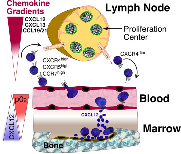
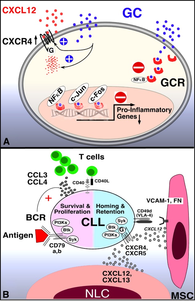
Chronic lymphocytic leukemia (CLL) cells proliferate in pseudofollicles within the lymphatic tissues, where signals from the microenvironment and BCR signaling drive the expansion of the CLL clone. Mobilization of tissue-resident cells into the blood removes CLL cells from this nurturing milieu and sensitizes them to cytotoxic drugs. This concept recently gained momentum after the clinical activity of kinase inhibitors that target BCR signaling (spleen tyrosine kinase, Bruton tyrosine kinase, PI3Kδ inhibitors) was established. Besides antiproliferative activity, these drugs cause CLL cell redistribution with rapid lymph node shrinkage, along with a transient surge in lymphocytosis, before inducing objective remissions. Inactivation of critical CLL homing mechanism (chemokine receptors, adhesion molecules), thwarting tissue retention and recirculation into the tissues, appears to be the basis for this striking clinical activity. This effect of BCR-signaling inhibitors resembles redistribution of CLL cells after glucocorticoids, described as early as in the 1940s. As such, we are witnessing a renaissance of the concept of leukemia cell redistribution in modern CLL therapy. Here, we review the molecular basis of CLL cell trafficking, homing, and redistribution and similarities between old and new drugs affecting these processes. In addition, we outline how these discoveries are changing our understanding of CLL biology and therapy.


Figures

Figure 1
Figure 1
CLL cell trafficking and tissue homing. CLL cells circulate in the peripheral blood, where they become attracted into the lymph nodes and bone marrow by chemokine gradients established by tissue stromal cells. Critical chemokines for lymph node homing are CXCL12, CXCL13, and CCL19/21, which bind to CXCR4, CXCR5, and CCR7 chemokine receptors on CLL cells, respectively. The CXCR4-CXCL12 axis is the predominant factor for marrow homing, and CXCL12 tissue expression is regulated by oxygen tension, as indicated by the triangles. Expression levels of CXCR4 on blood CLL cells can be used to distinguish CLL cells that are on their way into the tissues (CXCR4 high) versus CLL cells that recently have exited the tissues (CXCR4 dim). CLL adhesion molecules (integrins, selectins, CD44) co-operate with chemokine receptors during tissue homing. Pharmacologic inhibition of these homing mechanisms interferes with 2 distinct events: first, it leads to exit of tissue CLL cells into the blood, causing an increase in lymphocytosis. Second, this also causes inhibition of recirculation of blood CLL cells into the tissues.
Figure 2
Figure 2
Mechanism of GCs and BCR signaling inhibitors in CLL. (A) GCs inhibit transcription-dependent and -independent lymphocyte activation. Related to lymphocyte redistribution, GCs can up-regulate CXCR4 expression and signaling in normal T cells, thereby enhancing T-cell homing to the marrow. The mechanism of lymphocyte redistribution in CLL is currently unknown, but likely also is attributable to interference with homing mechanism. Besides effects on lymphocyte migration and homing, GCs bind to GCR in the cytosol, displacing heat-shock protein 90. GC-GCR complexes move into the nucleus, where they interfere with transcription. They also initiate transcription and translation of proteins, for example, of inhibitor of NF-κB (IκB). IκB then sequesters NF-κB. In addition, GC-GCR complexes can directly interact with NF-κB to suppress cytokine production. How these GCs mechanisms apply to CLL survival and proliferation is currently unknown. (B) Molecular interactions between CLL and stromal cells in the marrow and/or lymphoid tissue microenvironments and how these relate to BCR signaling and BCR-associated kinases (SYK, BTK, PI3K, modified after Figure 2 in Burger et al). BCR-associated kinases can influence CLL cell survival and proliferation (left) and CLL cell homing and retention in the tissues (right). Contact between CLL cells and NLC or mesenchymal stromal cells (MSC) is established and maintained by chemokine receptors and adhesion molecules expressed on CLL cells. NLCs express the chemokines CXCL12 and CXCL13, whereas MSCs predominantly express CXCL12. NLCs and MSCs attract CLL cells via the G-protein–coupled chemokine receptors CXCR4 and CXCR5, which are expressed at high levels on CLL cells. Integrins, particularly VLA-4 integrins (CD49d), expressed on the surface of CLL cells co-operate with chemokine receptors in establishing cell-cell adhesion through respective ligands on the stromal cells (VCAM-1 and fibronectin/FN). SYK, BTK, and PI3Ks are involved in chemokine receptor and adhesion molecule signaling in normal B cells and CLL cells. The clinical responses to small molecule antagonists to each of these kinases are characterized by “mobilization” of tissue-resident CLL cells into the blood, which indicates an important role of these kinases for CLL tissue homing and retention, as indicated in the diagram. Self and/or environmental antigens are considered a key factor in activation and expansion of the CLL clone. The nature and source of antigens and its mode of presentation to CLL cells are largely unknown. Stimulation of the BCR complex (BCR and CD79a,b) induces downstream signaling by recruitment and activation of SYK, BTK, and PI3Ks. Finally, BCR activation causes CLL cells to secrete high levels of the chemokines CCL3 and CCL4, which are potent T-cell attractants.
Figure 3
Figure 3
Transient lymphocytosis in CLL patients after treatment with the BTK inhibitor ibrutinib or GCs. (A) Trended ALC, hemoglobin levels (Hb), absolute neutrophil counts (ANC), and platelet counts (Plt) in 2 CLL patients (CLL #1, CLL #2) during continuous therapy with the BTK inhibitor ibrutinib at a dose of 420 MG daily. The horizontal axis shows the time of treatment. Please note the early, transient lymphocytosis, which peaked during the first weeks of therapy, then resolved, and both patients continue on therapy at 24+ months. (B) Effect of prednisone therapy on white cell counts in CLL patients (adapted from Shaw et al) is shown. In 16 of 18 patients a rapid increase in the total white cell count occurred during prednisone administration. This increase in the white count was noted early, the maximum occurring at 2 weeks, and could almost entirely be accounted for by an increase in lymphocytes. Subsequently the white cell count gradually decreased, reaching pretreatment values at the end of prednisone therapy and 2 months later were significantly below pretreatment values.

References

    1. Stein H, Bonk A, Tolksdorf G, Lennert K, Rodt H, Gerdes J. Immunohistologic analysis of the organization of normal lymphoid tissue and non-Hodgkin's lymphomas. J Histochem Cytochem. 1980;28(8):746–760. - PubMed
    1. Burger JA, Ghia P, Rosenwald A, Caligaris-Cappio F. The microenvironment in mature B-cell malignancies: a target for new treatment strategies. Blood. 2009;114(16):3367–3375. - PMC - PubMed
    1. Messmer BT, Messmer D, Allen SL, et al. In vivo measurements document the dynamic cellular kinetics of chronic lymphocytic leukemia B cells. J Clin Invest. 2005;115(3):755–764. - PMC - PubMed
    1. Tsukada N, Burger JA, Zvaifler NJ, Kipps TJ. Distinctive features of “nurselike” cells that differentiate in the context of chronic lymphocytic leukemia. Blood. 2002;99(3):1030–1037. - PubMed
    1. Burkle A, Niedermeier M, Schmitt-Graff A, Wierda WG, Keating MJ, Burger JA. Overexpression of the CXCR5 chemokine receptor, and its ligand, CXCL13 in B-cell chronic lymphocytic leukemia. Blood. 2007;110(9):3316–3325. - PubMed

Publication types

MeSH terms