Review
. 2013 Oct;84(10):1161-70.
doi: 10.1136/jnnp-2012-304019. Epub 2012 Dec 21.

Transcranial magnetic stimulation and amyotrophic lateral sclerosis: pathophysiological insights

Affiliations
Free PMC article
Review

Transcranial magnetic stimulation and amyotrophic lateral sclerosis: pathophysiological insights

Steve Vucic et al. J Neurol Neurosurg Psychiatry. 2013 Oct.
Free PMC article

Abstract

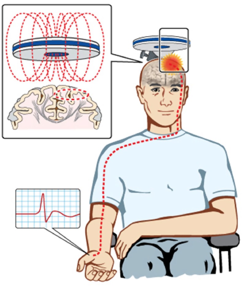
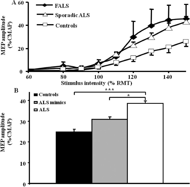
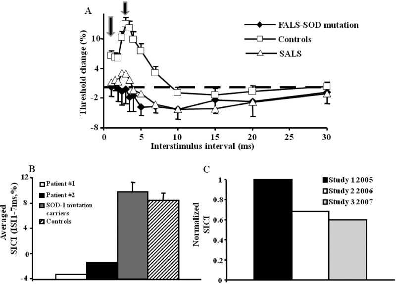
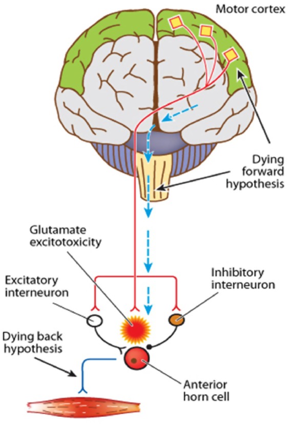
Amyotrophic lateral sclerosis (ALS) is a rapidly progressive neurodegenerative disorder of the motor neurons in the motor cortex, brainstem and spinal cord. A combination of upper and lower motor neuron dysfunction comprises the clinical ALS phenotype. Although the ALS phenotype was first observed by Charcot over 100 years ago, the site of ALS onset and the pathophysiological mechanisms underlying the development of motor neuron degeneration remain to be elucidated. Transcranial magnetic stimulation (TMS) enables non-invasive assessment of the functional integrity of the motor cortex and its corticomotoneuronal projections. To date, TMS studies have established motor cortical and corticospinal dysfunction in ALS, with cortical hyperexcitability being an early feature in sporadic forms of ALS and preceding the clinical onset of familial ALS. Taken together, a central origin of ALS is supported by TMS studies, with an anterograde transsynaptic mechanism implicated in ALS pathogenesis. Of further relevance, TMS techniques reliably distinguish ALS from mimic disorders, despite a compatible peripheral disease burden, thereby suggesting a potential diagnostic utility of TMS in ALS. This review will focus on the mechanisms underlying the generation of TMS measures used in assessment of cortical excitability, the contribution of TMS in enhancing the understanding of ALS pathophysiology and the potential diagnostic utility of TMS techniques in ALS.

Keywords: ALS; EMG.


Figures

Figure 1
Figure 1
Transcranial magnetic stimulation excites a network of neurons in the underlying motor cortex with motor evoked potentials recorded over the contralateral abductor pollicis brevis muscle. The motor cortex is preferentially stimulated when the current flows in a posterior–anterior direction within the motor cortex.
Figure 2
Figure 2
(A) The motor evoked potential (MEP) amplitude, expressed as a percentage of compound muscle action potential (CMAP) response, is significantly increased in sporadic amyotrophic lateral sclerosis (ALS) and familial ALS (FALS) when compared with healthy controls. (B) The MEP amplitude is significantly increased in ALS when compared with pathological and healthy controls, thereby distinguishing ALS from ALS mimic disorders.* p<0.05; ***p<0.001. RMT, resting motor threshold.
Figure 3
Figure 3
(A) Short interval intracortical inhibition (SICI), defined as the stimulus intensity required to maintain a target motor evoked potential of 0.2 mV, as assessed by the threshold tracking transcranial magnetic stimulation technique. Intracortical inhibition is illustrated by an increase in the conditioned test stimulus intensity required to track the target response, while intracortical facilitation is indicated by a reduction in test stimulus intensity. In healthy controls, SICI develops between interstimulus intervals (ISI) of 1 and 7 ms, with two peaks evident at 1 and 3 ms as indicated by the arrows. Intracortical facilitation developed between ISIs of 10 and 30 ms. SICI is significantly reduced in both sporadic amyotrophic lateral sclerosis (SALS) and familial amyotrophic lateral sclerosis (FALS). (B) Averaged SICI, between ISI 1 and 7 ms, was reduced in two presymptomatic superoxide dismutase-1 (SOD-1) mutation carriers 6 months prior to the development of ALS. (C) Normalised SICI, expressed as a fraction of the SICI value measured at the first study, was reduced 8 months prior to development of ALS in a third presymptomatic SOD-1 mutation carrier.
Figure 4
Figure 4
The dying forward and dying back hypothesis of amyotrophic lateral sclerosis (ALS). The ‘dying forward’ hypothesis proposed that ALS was primarily a disorder of the corticomotoneurons (highlighted in red), with anterior horn cell degeneration mediated via an anterograde glutamate-mediated excitotoxic process. In contrast, the dying back hypothesis proposes that ALS begins within the muscle or neuromuscular junction, with pathogens retrogradely transported from the neuromuscular junction to the cell body where these pathogens may exert their deleterious effects.

References

    1. Charcot J, Joffroy A. Deux cas d'atrophie musculaire progressive avec lesion de la substance grise et des faisceaux antero-lateraux de la moelle epiniere. Arch Physiol Neurol Pathol 1869;2:744–54
    1. Kiernan MC, Vucic S, Cheah BC, et al. Amyotrophic lateral sclerosis. Lancet 2011;377:942–55 - PubMed
    1. Vucic S, Burke D, Kiernan MC. Diagnosis of motor neuron disease. In: Kiernan MC. ed. The motor neuron disease handbook. Sydney: Australasian Medical Publishing Company Limited, 2007:89–115
    1. Winhammar JM, Rowe DB, Henderson RD, et al. Assessment of disease progression in motor neuron disease. Lancet Neurology 2005;4:229–38 - PubMed
    1. Ravits J, Paul P, Jorg C. Focality of upper and lower motor neuron degeneration at the clinical onset of ALS. Neurology 2007;68:1571–5 - PubMed

Publication types

MeSH terms