. 2013 Feb 15;126(Pt 4):953-65.
doi: 10.1242/jcs.116426. Epub 2012 Dec 21.

PDGFRα signaling in the primary cilium regulates NHE1-dependent fibroblast migration via coordinated differential activity of MEK1/2-ERK1/2-p90RSK and AKT signaling pathways

Affiliations

PDGFRα signaling in the primary cilium regulates NHE1-dependent fibroblast migration via coordinated differential activity of MEK1/2-ERK1/2-p90RSK and AKT signaling pathways

Ditte L Clement et al. J Cell Sci. .

Abstract

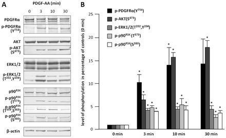
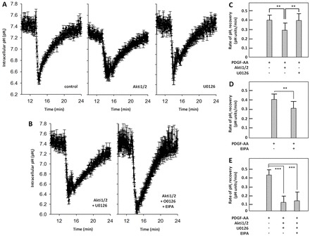
In fibroblasts, platelet-derived growth factor receptor alpha (PDGFRα) is upregulated during growth arrest and compartmentalized to the primary cilium. PDGF-AA mediated activation of the dimerized ciliary receptor produces a phosphorylation cascade through the PI3K-AKT and MEK1/2-ERK1/2 pathways leading to the activation of the Na(+)/H(+) exchanger, NHE1, cytoplasmic alkalinization and actin nucleation at the lamellipodium that supports directional cell migration. We here show that AKT and MEK1/2-ERK1/2-p90(RSK) inhibition reduced PDGF-AA-induced cell migration by distinct mechanisms: AKT inhibition reduced NHE1 activity by blocking the translocation of NHE1 to the cell membrane. MEK1/2 inhibition did not affect NHE1 activity but influenced NHE1 localization, causing NHE1 to localize discontinuously in patches along the plasma membrane, rather than preferentially at the lamellipodium. We also provide direct evidence of NHE1 translocation through the cytoplasm to the leading edge. In conclusion, signals initiated at the primary cilium through the PDGFRαα cascade reorganize the cytoskeleton to regulate cell migration differentially through the AKT and the MEK1/2-ERK1/2-p90(RSK) pathways. The AKT pathway is necessary for initiation of NHE1 translocation, presumably in vesicles, to the leading edge and for its activation. In contrast, the MEK1/2-ERK1/2-p90(RSK) pathway controls the spatial organization of NHE1 translocation and incorporation, and therefore specifies the direction of the leading edge formation.


Figures

Fig. 1.
Fig. 1.
Signaling pathways activated by PDGF-AA in NIH3T3 cells. (A) WB analysis of NIH3T3 cells after 24 h serum deprivation and stimulation with 25 ng/ml PDGF-AA for 0, 3, 10 or 30 min. Antibodies used were directed against total PDGFRα, Y754-phosphorylated PDGFRα, AKT, S473-phosphorylated AKT, ERK1/2, T202/Y204-phosphorylated ERK1/2, p90RSK, T573-phosphorylated p90RSK, T359/S363-phosphorylated p90RSK and S380-phosphorylated p90RSK; β-actin was used as a loading control. (B) Quantification of WB analysis and ANOVA (n≧3). The values at 0 min of stimulation were set to 1 and those at 3, 10 and 30 min of stimulation are the percentage phosphorylation compared to non-stimulated cells. *Significantly different from the respective control (P<0.05).
Fig. 2.
Fig. 2.
p90RSK is activated by MEK1/2-ERK1/2 at the primary cilium. (A) WB analysis of the effects of the AKT inhibitor (Akti1/2: 0.1, 0.3, 1, 3, 10 µM, 1 h) and the MEK1/2 inhibitor (U0126: 0.3, 1, 3, 10 µM, 1 h) on PDGF-AA-mediated signal transduction in growth-arrested NIH3T3 cells. PDGF-AA was added at 25 ng/ml for 10 min. (B) Quantification of WB analysis and ANOVA (n = 3). The inhibition of AKT by Akti1/2, and ERK1/2 and RSK by U0126 is considered extremely significant (***P<0.001). (C–F) IFM analysis of primary cilia after 24 h of serum starvation. ‘PDGF-AA’ indicates that cells were stimulated with the ligand for 10 min (25 ng/ml). Phospho-MEK1/2 (pMEK1/2), p90RSK and phospho-p90RSK (p-p90RSK) are green; anti-acetylated α-tubulin (Ac-tub; in red) marks stable microtubules, including primary cilia (arrows). Nuclei were stained with DAPI (blue). (G,H) Cells were stimulated with PDGF-AA (25 ng/ml for 10 min) followed by IFM analysis to confirm localization of phospho-p90RSK at the ciliary base. Primary cilia are marked with Ac-tub (blue, arrows) and centrosomes with anti-EB3 (red, asterisks); p-p90RSK is green. (I) Cells without and with blocking peptide against p-p90RSK(T573). (J) Cells were pre-incubated for 1 h with either 10 µM U0126 or Akti1/2 followed by stimulation with PDGF-AA (25 ng/ml for 10 min) and IFM analysis. Primary cilia are marked with Ac-tub (red, arrows); p-p90RSK is green (open arrows). The lower row of panels show shifted images, where p-p90RSK is shifted to the right. (K) Quantification of relative p-p90RSK fluorescence levels at the base of the cilia, from the cells shown in J. Reduced localization of p-p90RSK fluorescence at the base of the cilia in the presence of U0126 is extremely significant (***P<0.001; ANOVA; n = 30).
Fig. 3.
Fig. 3.
PDGF-AA-mediated activation of the PI3K-AKT and MEK1/2-ERK1/2-p90RSK pathways is abolished at stunted primary cilia in Tg737orpk MEFs. (A) WB analysis after 24 h of serum deprivation of wt and Tg737orpk MEF cells stimulated with or without PDGF-AA and PDGF-BB. Control cells are incubated with DMSO. (B) Quantification of WB analysis and ANOVA (n = 3). The inhibition of AKT by Akti1/2, and ERK1/2 and p90RSK by U0126 is either very significant (**P<0.01) or extremely significant (***P<0.001). (C) IFM analysis of wt and Tg737orpk MEFs with antibodies against anti-acetylated α-tubulin (Ac-tub, red), which marks stable microtubules, including primary cilia (arrows) and p-p90RSK(T359/S363) (green). Nuclei were stained with DAPI (blue). Upper right inserts show magnifications of the ciliary base localization of p-p90RSK(T359/S363). Lower right inserts show the ciliary base localization of p-p90RSK(T573) (green). Open arrows indicate stunted cilia in Tg737orpk MEFs. Asterisks mark the base of a cilium. (D) Quantification of relative p-p90RSK fluorescence levels at the base of the cilia from data shown in Fig. 2C. Reduced localization of p-p90RSK fluorescence at the ciliary base in Tg737orpk MEFs is extremely significant (***P<0.001; ANOVA; n = 30).
Fig. 4.
Fig. 4.
Effects of PDGF-AA, Akti1/2, U0126 and EIPA in wound-healing and localization of NHE1 in growth-arrested NIH3T3 fibroblasts. (A) Wound healing assay trajectories of growth-arrested NIH3T3 cells. Each line represents the migration of one cell within a 4 h period. The red circles illustrate the mean translocation of the cells. (B) Translocation in the absence or presence of inhibitor (10 µM) and PDGF-AA (25 ng/ml) as indicated (ANOVA: *P<0.05; ***P<0.001; n = 3). (C) IFM analysis of cell culture with scratch after 24 h of serum starvation. Anti-α-tubulin (Tub, red) marks microtubules, the actin cytoskeleton is stained with phalloidin (F-actin, blue) and NHE1 is stained with anti-NHE1 (green, bold arrows). Differential interference contrast (DIC) images show the three-dimensional appearance. Open arrows indicate direction of migration into the scratch. The lowest row of panels shows magnifications of the boxed areas in the fourth row of merged images. The dotted lines indicate the surface of the cells facing the scratch.
Fig. 5.
Fig. 5.
Effects of Akti1/2 and U0126 on focal contact formation and NHE1 localization during wound healing in growth-arrested NIH3T3 fibroblasts. IFM analysis of NIH3T3 cell culture with scratch after 24 h of serum starvation with antibodies against vinculin (red) and NHE1 (green, bold arrows); the actin cytoskeleton is stained with phalloidin (F-actin, blue). DIC images show the three-dimensional appearance. Open arrows indicate direction of migration into the scratch. The lowest row of panels shows magnifications of the boxed areas in the fourth row of merged images. The dotted lines indicate the surface of the cells facing the scratch.
Fig. 6.
Fig. 6.
Translocation of NHE1–GFP to the leading edge of migrating MEFs. (A) WB analysis of NHE1–GFP expression in non-transfected and stably expressing MEFs in cycling cells (+ serum) and in growth-arrested cells (− serum). (B) Effects of Akti1/2 and U0126 in scratch assays on localization of NHE1–GFP (green) in growth-arrested MEFs stimulated with PDGF-AA by IFM analysis. The NHE1–GFP signal was increased with anti-GFP. Open arrows indicate direction of movement into the scratch. Solid arrows indicate NHE1–GFP localization at the cell edges. The analysis was carried out in more than 30 transfected cells with similar result.
Fig. 7.
Fig. 7.
Effects of MEK1/2, AKT and NHE1 inhibitors on intracellular pH recovery during growth arrest in NIH3T3 cells. (A,B) Tracings of pHi recovery measurements after acidification and stimulation with PDGF-AA (25 ng/ml) and in the absence or (A) presence of Akti1/2 (10 µM) or U0126 (10 µM), and (B) Akti1/2+U0126 or Akti1/2+U0126+EIPA. (C–E) Summary of the pHi recovery rates under the conditions shown. The rate of pHi recovery (in pH units/minute) was calculated from the slope of the initial linear part of the curve after NH4Cl removal. Data were analyzed using ANOVA: **P<0.01 ***P<0.001 (n = 6).

References

    1. Anjum R., Blenis J. (2008). The RSK family of kinases: emerging roles in cellular signalling. Nat. Rev. Mol. Cell Biol. 9, 747–758. 10.1038/nrm2509 - DOI - PubMed
    1. Boedtkjer E., Bunch L., Pedersen S. F. (2012). Physiology, pharmacology and pathophysiology of the pH regulatory transport proteins NHE1 and NBCn1: similarities, differences, and implications for cancer therapy. Curr. Pharm. Des. 18, 1345–1371. - PubMed
    1. Boron W. F. (2004). Regulation of intracellular pH. Adv. Physiol. Educ. 28, 160–179. - PubMed
    1. Boyarsky G., Ganz M. B., Sterzel R. B., Boron W. F. (1988). pH regulation in single glomerular mesangial cells. II. Na+-dependent and -independent Cl(-)-HCO3- exchangers. Am. J. Physiol. 255, C857–C869. - PubMed
    1. Chen X. W., Saltiel A. R. (2011). Ral's engagement with the exocyst: breaking up is hard to do. Cell Cycle 10, 2299–2304. 10.4161/cc.10.14.16524 - DOI - PubMed

Publication types

MeSH terms

Substances