Different distribution of neuromedin S and its mRNA in the rat brain: NMS peptide is present not only in the hypothalamus as the mRNA, but also in the brainstem
- PMID: 23264767
- PMCID: PMC3524995
- DOI: 10.3389/fendo.2012.00152
Different distribution of neuromedin S and its mRNA in the rat brain: NMS peptide is present not only in the hypothalamus as the mRNA, but also in the brainstem
Abstract
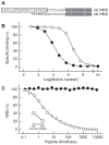
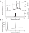
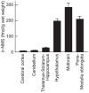
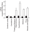
Neuromedin S (NMS) is a neuropeptide identified as another endogenous ligand for two orphan G protein-coupled receptors, FM-3/GPR66 and FM-4/TGR-1, which have also been identified as types 1 and 2 receptors for neuromedin U structurally related to NMS. Although expression of NMS mRNA is found mainly in the brain, spleen, and testis, the distribution of its peptide has not yet been investigated. Using a newly prepared antiserum, we developed a highly sensitive radioimmunoassay for rat NMS. NMS peptide was clearly detected in the rat brain at a concentration of 68.3 ± 3.4 fmol/g wet weight, but it was hardly detected in the spleen and testis. A high content of NMS peptide was found in the hypothalamus, midbrain, and pons-medulla oblongata, whereas abundant expression of NMS mRNA was detected only in the hypothalamus. These differing distributions of the mRNA and peptide suggest that nerve fibers originating from hypothalamic NMS neurons project into the midbrain, pons, or medulla oblongata. In addition, abundant expression of type 2 receptor mRNA was detected not only in the hypothalamus, but also in the midbrain and pons-medulla oblongata. These results suggest novel, unknown physiological roles of NMS within the brainstem.
Keywords: brain; brainstem; neuromedin S; neuropeptide; peptide distribution; radioimmunoassay.
Figures


Similar articles
-
Neuromedin s is a novel anorexigenic hormone.Endocrinology. 2005 Oct;146(10):4217-23. doi: 10.1210/en.2005-0107. Epub 2005 Jun 23. Endocrinology. 2005. PMID: 15976061
-
Neuromedin S: discovery and functions.Results Probl Cell Differ. 2008;46:201-12. doi: 10.1007/400_2007_054. Results Probl Cell Differ. 2008. PMID: 18214396 Review.
-
Identification and functional analysis of a novel ligand for G protein-coupled receptor, Neuromedin S.Regul Pept. 2008 Jan 10;145(1-3):37-41. doi: 10.1016/j.regpep.2007.08.013. Epub 2007 Aug 24. Regul Pept. 2008. PMID: 17870195
-
Identification of neuromedin S and its possible role in the mammalian circadian oscillator system.EMBO J. 2005 Jan 26;24(2):325-35. doi: 10.1038/sj.emboj.7600526. Epub 2005 Jan 6. EMBO J. 2005. PMID: 15635449 Free PMC article.
-
Emerging pharmacology and physiology of neuromedin U and the structurally related peptide neuromedin S.Br J Pharmacol. 2009 Sep;158(1):87-103. doi: 10.1111/j.1476-5381.2009.00252.x. Epub 2009 Jun 10. Br J Pharmacol. 2009. PMID: 19519756 Free PMC article. Review.
Cited by
-
Identification of neuromedin U precursor-related peptide and its possible role in the regulation of prolactin release.Sci Rep. 2017 Sep 5;7(1):10468. doi: 10.1038/s41598-017-10319-9. Sci Rep. 2017. PMID: 28874765 Free PMC article.
-
Neuromedin S Regulates Steroidogenesis through Maintaining Mitochondrial Morphology and Function via NMUR2 in Goat Ovarian Granulosa Cells.Int J Mol Sci. 2022 Nov 2;23(21):13402. doi: 10.3390/ijms232113402. Int J Mol Sci. 2022. PMID: 36362185 Free PMC article.
-
Neuromedins NMU and NMS: An Updated Overview of Their Functions.Front Endocrinol (Lausanne). 2021 Jul 1;12:713961. doi: 10.3389/fendo.2021.713961. eCollection 2021. Front Endocrinol (Lausanne). 2021. PMID: 34276571 Free PMC article. Review.
References
-
- Aston-Jones G., Chen S., Zhu Y., Oshinsky M. L. (2001). A neural circuit for circadian regulation of arousal. Nat. Neurosci. 4 732–738 - PubMed
-
- Brighton P. J., Szekeres P. G., Willars G. B. (2004). Neuromedin U and its receptors: structure, function, and physiological roles. Pharmacol. Rev. 56 231–248 - PubMed
-
- Fujii R., Hosoya M., Fukusumi S., Kawamata Y., Habata Y., Hinuma S., et al. (2000). Identification of neuromedin U as the cognate ligand of the orphan G protein-coupled receptor FM-3. J. Biol. Chem. 275 21068–21074 - PubMed
LinkOut - more resources
Full Text Sources

