

Trypanosoma cruzi extracellular amastigotes and host cell signaling: more pieces to the puzzle
- PMID: 23264776
- PMCID: PMC3525110
- DOI: 10.3389/fimmu.2012.00363
Trypanosoma cruzi extracellular amastigotes and host cell signaling: more pieces to the puzzle
Abstract
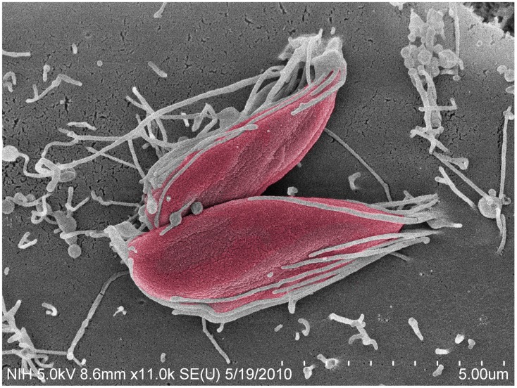
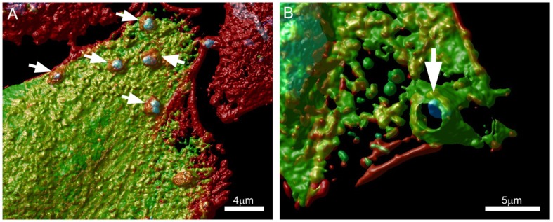
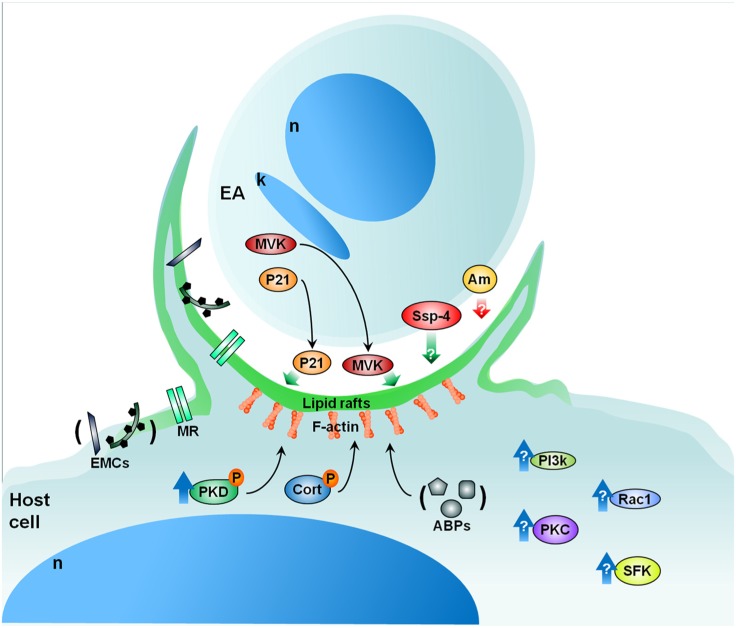
Among the different infective stages that Trypanosoma cruzi employs to invade cells, extracellular amastigotes (EAs) have recently gained attention by our group. This is true primarily because these amastigotes are able to infect cultured cells and animals, establishing a sustainable infective cycle. EAs are thus an excellent means of adaptation and survival for T. cruzi, whose different infective stages each utilize unique mechanisms for attachment and penetration. Here we discuss some features of host cell invasion by EAs and the associated host cell signaling events that occur as part of the process.
Keywords: cell invasion; extracellular amastigotes; mevalonate kinase; protein kinase D; signaling.
Figures
References
-
- Abrahamsohn I. A., Katzin A. M., Milder R. V. (1983). A method for isolating Trypanosoma cruzi amastigotes from spleen and liver using two-step discontinuous gradient centrifugation. J. Parasitol. 69 437–439 - PubMed
-
- Andreoli W. K., Mortara R. A. (2003). Acidification modulates the traffic of Trypanosoma cruzi trypomastigotes in Vero cells harboring Coxiella burnetti vacuoles. Int. J. Parasitol. 33 185–197 - PubMed
-
- Andrews N. W., Hong K. S., Robbins E., Nussenzweig V. (1987). Stage specific surface antigens expressed during the morphogenesis of vertebrate forms of Trypanosoma cruzi. Exp. Parasitol. 64 474–484 - PubMed
-
- Bambino-Medeiros R., Oliveira F. O., Calvet C. M., Vicente D., Toma L., Krieger M. A., et al. (2011). Involvement of host cell heparan sulfate proteoglycan in Trypanosoma cruzi amastigote attachment and invasion. Parasitology 138 593–601 - PubMed
-
- Barrias E. S., Dutra J. M., De Souza W., Carvalho T. M. (2007). Participation of macrophage membrane rafts in Trypanosoma cruzi invasion process. Biochem. Biophys. Res. Commun. 2363 828–834 - PubMed
LinkOut - more resources
Full Text Sources