

Pattern Visual Evoked Potentials in Patients with Type II Diabetes Mellitus
- PMID: 23264865
- PMCID: PMC3520591
Pattern Visual Evoked Potentials in Patients with Type II Diabetes Mellitus
Abstract
Purpose: To evaluate cortical and retinal activity by pattern visual evoked potentials (PVEP) in patients with type II diabetes mellitus.
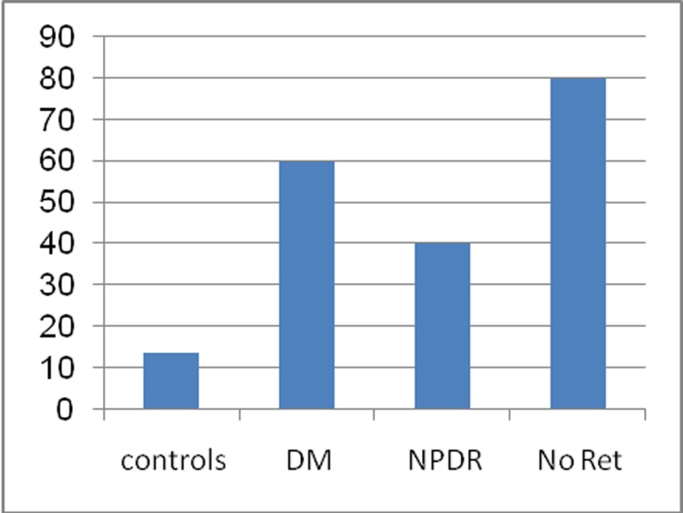
Methods: PVEP was recorded in 40 diabetic patients including 20 subjects with non-proliferative diabetic retinopathy (NPDR) and 20 others without any retinopathy on fundus photography, and compared to 40 age- and sex-matched normal non-diabetic controls.
Results: P100 wave latency was significantly longer in diabetic patients as compared to normal controls (P<0.001); both diabetic subjects without retinopathy and those with NPDR had significantly longer P100 latency than controls (P<0.001 for both comparisons). There was significant reduction in N75 (P=0.037) and P100 (P=0.001) amplitudes in diabetic subjects. No correlation was observed between VEP amplitude or wave latency, and the level of glycemia or duration of diabetes mellitus.
Conclusion: Increased PVEP latency may be a sign of retinal ganglion cell damage which takes place before the appearance of the first ophthalmoscopically detectable signs of diabetic retinopathy. PVEP may be considered as a method for detecting prediabetic retinopathy and has the potential to reduce diabetic complications.
Keywords: Diabetic Retinopathy; Pattern VEP; Type II Diabetes Mellitus.
Figures
Similar articles
-
Visual Evoked Potentials for the Detection of Diabetic Retinal Neuropathy.Int J Mol Sci. 2023 Apr 17;24(8):7361. doi: 10.3390/ijms24087361. Int J Mol Sci. 2023. PMID: 37108524 Free PMC article. Review.
-
Utility of Visual Evoked Potentials (VEPs) study in the evaluation of visual pathway dysfunction in diabetics without retinopathy: correlations with diabetic peripheral neuropathy and other clinical findings.Rom J Ophthalmol. 2024 Apr-Jun;68(2):114-121. doi: 10.22336/rjo.2024.22. Rom J Ophthalmol. 2024. PMID: 39006331 Free PMC article.
-
Visual evoked potential can be used to detect a prediabetic form of diabetic retinopathy in patients with diabetes mellitus type I.Coll Antropol. 2010 Jun;34(2):525-9. Coll Antropol. 2010. PMID: 20698126
-
Evaluation of visual pathways using visual evoked potential in patients with diabetic retinopathy.Rom J Ophthalmol. 2019 Oct-Dec;63(4):367-371. Rom J Ophthalmol. 2019. PMID: 31915735 Free PMC article.
-
Visual Evoked Potential to Assess Retinopathy in Gestational Diabetes Mellitus.Can J Diabetes. 2016 Apr;40(2):131-4. doi: 10.1016/j.jcjd.2015.08.009. Epub 2015 Dec 24. Can J Diabetes. 2016. PMID: 26724915
Cited by
-
Electrophysiological changes in optic neuropathy of streptozotocin induced diabetic rats.J Med Life. 2013 Sep 15;6(3):340-8. Epub 2013 Sep 25. J Med Life. 2013. PMID: 24155786 Free PMC article.
-
Neuroinflammation and oxidative stress act in concert to promote neurodegeneration in the diabetic retina and optic nerve: galectin-3 participation.Neural Regen Res. 2020 Apr;15(4):625-635. doi: 10.4103/1673-5374.266910. Neural Regen Res. 2020. PMID: 31638084 Free PMC article. Review.
-
Visual Evoked Potentials for the Detection of Diabetic Retinal Neuropathy.Int J Mol Sci. 2023 Apr 17;24(8):7361. doi: 10.3390/ijms24087361. Int J Mol Sci. 2023. PMID: 37108524 Free PMC article. Review.
-
Utility of Visual Evoked Potentials (VEPs) study in the evaluation of visual pathway dysfunction in diabetics without retinopathy: correlations with diabetic peripheral neuropathy and other clinical findings.Rom J Ophthalmol. 2024 Apr-Jun;68(2):114-121. doi: 10.22336/rjo.2024.22. Rom J Ophthalmol. 2024. PMID: 39006331 Free PMC article.
-
Cerebral perfusion alterations in type 2 diabetes and its relation to insulin resistance and cognitive dysfunction.Brain Imaging Behav. 2017 Oct;11(5):1248-1257. doi: 10.1007/s11682-016-9583-9. Brain Imaging Behav. 2017. PMID: 27714551 Free PMC article.
References
-
- Silink M. IDF Diabetes Atlas. 4th. Brussels: International Diabetes Federation; 2009.
-
- Biessels GJ, Koffeman A, Scheltens P. Diabetes and cognitive impairment. Clinical diagnosis and brain imaging in patients attending a memory clinic. J Neurol. 2006;253:477–482. - PubMed
-
- Algan M, Ziegler O, Gehin P, Got I, Raspiller A, Weber M, et al. Visual evoked potentials in diabetic patients. Diabetes Care. 1989;12:227–229. - PubMed
-
- Gregori B, Galie E, Pro S, Clementi A, Accornero N. Luminance and chromatic visual evoked potentials in type I and II diabetes: relationships with peripheral neuropathy. Neurol Sci. 2006;27:323–327. - PubMed
-
- Locke S. The nervous system and diabetes. In: Marble A, et al., editors. Joslin's Diabetes Mellitus. 11th ed. Philadelphia: Lea and Febiger; 1971. pp. 562–564.
LinkOut - more resources
Full Text Sources