Expression of antigen tf and galectin-3 in fibroadenoma
- PMID: 23265237
- PMCID: PMC3532378
- DOI: 10.1186/1756-0500-5-694
Expression of antigen tf and galectin-3 in fibroadenoma
Abstract
Background: Fibroadenomas are benign human breast tumors, characterized by proliferation of epithelial and stromal components of the terminal ductal unit. They may grow, regress or remain unchanged, as the hormonal environment of the patient changes. Expression of antigen TF in mucin or mucin-type glycoproteins and of galectin-3 seems to contribute to proliferation and transformations events; their expression has been reported in ductal breast cancer and in aggressive tumors.
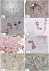
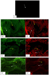
Findings: Lectin histochemistry, immunohistochemistry, and immunofluorescence were used to examine the expression and distribution of antigen TF and galectin-3. We used lectins from Arachis hypogaea, Artocarpus integrifolia, and Amaranthus lecuocarpus to evaluate TF expression and a monoclonal antibody to evaluate galectin-3 expression. We used paraffin-embedded blocks from 10 breast tissues diagnosed with fibroadenoma and as control 10 healthy tissue samples. Histochemical and immunofluorescence analysis showed positive expression of galectin-3 in fibroadenoma tissue, mainly in stroma, weak interaction in ducts was observed; whereas, in healthy tissue samples the staining was also weak in ducts. Lectins from A. leucocarpus and A. integrifolia specificaly recognized ducts in healthy breast samples, whereas the lectin from A. hypogaea recognized ducts and stroma. In fibroadenoma tissue, the lectins from A. integrifolia, A. Hypogaea, and A. leucocarpus recognized mainly ducts.
Conclusions: Our results suggest that expression of antigen TF and galectin-3 seems to participate in fibroadenoma development.
Figures


Similar articles
-
O-glycosylation expression in fibroadenoma.Prep Biochem Biotechnol. 2010;40(1):1-12. doi: 10.1080/10826060903386071. Prep Biochem Biotechnol. 2010. PMID: 20024790
-
Identification of galectin-3 and mucin-type O-glycans in breast cancer and its metastasis to brain.Cancer Invest. 2008 Jul;26(6):615-23. doi: 10.1080/07357900701837051. Cancer Invest. 2008. PMID: 18584353
-
Mammary fibroadenoma and some phyllodes tumour stroma are composed of CD34+ fibroblasts and factor XIIIa+ dendrophages.Histopathology. 1996 Nov;29(5):411-9. doi: 10.1046/j.1365-2559.1996.d01-510.x. Histopathology. 1996. PMID: 8951485
-
Analysis of CD83 antigen expression in human breast fibroadenoma and adjacent tissue.Sao Paulo Med J. 2011 Dec;129(6):402-9. doi: 10.1590/s1516-31802011000600006. Sao Paulo Med J. 2011. PMID: 22249796 Free PMC article.
-
Lectin-epithelial interactions in the human colon.Biochem Soc Trans. 2008 Dec;36(Pt 6):1482-6. doi: 10.1042/BST0361482. Biochem Soc Trans. 2008. PMID: 19021580 Review.
References
-
- Dixon JM. Cystic disease and fibroadenoma of the breast: natural history and relation to breast cancer risk. Br Med Bull. 1991;47:258–271. - PubMed
-
- Martin PM, Kuttenn F, Serment H, Mauvais-Jarvis P. Studies on clinical, hormonal and pathological correlations in breast fibroadenomas. J Steroid Biochem. 1978;912:1251–1255. - PubMed
Publication types
MeSH terms
Substances
LinkOut - more resources
Full Text Sources
Other Literature Sources
Medical
Miscellaneous

