. 2013 Oct;8(10):1601-9.
doi: 10.2217/nnm.12.165. Epub 2012 Dec 24.

Gold nanoparticle imaging and radiotherapy of brain tumors in mice

Affiliations

Gold nanoparticle imaging and radiotherapy of brain tumors in mice

James F Hainfeld et al. Nanomedicine (Lond). 2013 Oct.

Abstract

Aim: To test intravenously injected gold nanoparticles for x-ray imaging and radiotherapy enhancement of large, imminently lethal, intracerebral malignant gliomas.

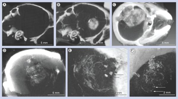
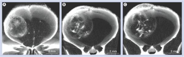
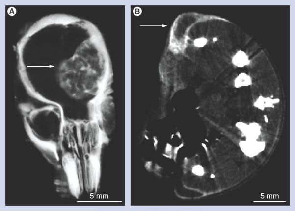
Materials & methods: Gold nanoparticles approximately 11 nm in size were injected intravenously and brains imaged using microcomputed tomography. A total of 15 h after an intravenous dose of 4 g Au/kg was administered, brains were irradiated with 30 Gy 100 kVp x-rays.

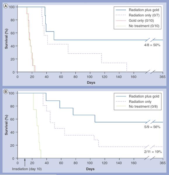
Results: Gold uptake gave a 19:1 tumor to normal brain ratio with 1.5% w/w gold in tumor, calculated to increase local radiation dose by approximately 300%. Mice receiving gold and radiation (30 Gy) demonstrated 50% long term (>1 year) tumor-free survival, whereas all mice receiving radiation only died.

Conclusion: Intravenously injected gold nanoparticles cross the blood-tumor barrier, but are largely blocked by the normal blood-brain barrier, enabling high-resolution computed tomography tumor imaging. Gold radiation enhancement significantly improved long-term survival compared with radiotherapy alone. This approach holds promise to improve therapy of human brain tumors and other cancers.


Figures

Figure 1
Figure 1. Irradiation and tumor dissection
(A) Irradiation setup showing mouse with a lead collimator. (B) Mouse brain and glioma at necropsy 10 days after tumor initiation and 4 h after gold nanoparticle intravenous injection (4 g Au/kg). The tumor (curved arrow) was removed (place of removal: straight arrow). The removed tumor was black compared with normal brain tissue due to gold nanoparticle uptake.
Figure 2
Figure 2. Microcomputed tomography imaging of brain tumors after intravenous gold nanoparticle injection
(A–C) Live mouse microcomputed tomography images of brain tumors 9 days postimplantation and 15 h after intravenous (iv.) gold nanoparticle (AuNP) injection. (A & B) Same mouse (A) before and (B) 15 h after iv. injection (4 g Au/kg). (C) Larger tumor imaged after iv. injection of 1.7 g Au/kg. (D–F) Live mouse microcomputed tomography images of a typical brain tumor 9 days postimplantation and 1 h after iv. AuNP injection (1.7 g Au/kg). (D) Individual blood vessels in the tumor could be discerned. (E) Focal spots of gold were also observed (arrows). (F) AuNP leakage was very irregular in some vessels (arrows).
Figure 3
Figure 3. Tumor imaging time course over 8 days after intravenous gold nanoparticle injection
The same live mouse microcomputed tomography tumor images taken (A) 1 day, (B) 3 days and (C) 8 days after intravenous injection of gold nanoparticles (2 g Au/kg). Note how the gold expands with the tumor boundary, but becomes more punctuate and condensed internally. (C) Tumor is also infiltrating the scalp.
Figure 4
Figure 4. Microcomputed tomography sections comparing glioma versus subcutaneous tumor gold nanoparticle distribution after intravenous injection
Thin microcomputed tomography sections through the center of tumors of live mice bearing (A) an orthotopic brain tumor or (B) two subcutaneous tumors, one on each leg, see arrows. (A) Note how the gold nanoparticles (white contrast) are distributed throughout the brain tumor, even in its center, whereas (B) the subcutaneous tumors have gold nanoparticles predominantly at their periphery.
Figure 5
Figure 5. Kaplan-Meier survival graphs of mice with brain tumors after various treatments
(A) Groups: radiation only (30 Gy); radiation (30 Gy) plus intravenous (iv.) gold nanparticles (AuNPs; 4 g Au/kg); no treatment; and iv. AuNPs only (4 g Au/kg). Long-term survival (>365 days) was 50% in the gold plus radiation group (n = 8), and 0% in the untreated (n = 10), gold-only (n = 10) and radiation-only (n = 7) groups. (B) Repeat of experiment shown in (A), except using 35 Gy instead of 30 Gy. Groups: no treatment; radiation only (35 Gy); and radiation (35 Gy) plus iv. AuNPs (4 g Au/kg). Long-term survival (>365 days) was 56% in the gold plus radiation group (n = 9), 18% in the radiation-only group (n = 11) and 0% in the untreated group (n = 8).

References

    1. Beier D, Schulz JB, Beier CP. Chemoresistance of glioblastoma cancer stem cells: much more complex than expected. Mol. Canc. 2011;10:128. - PMC - PubMed
    1. Mrugala MM, Adair JE, Kiem HP. Outside the box: novel therapeutic strategies for glioblastoma. Cancer J. 2012;18(1):51–58. - PubMed
    1. Liu HL, Yang HW, Hua MY, Wei KC. Enhanced therapeutic agent delivery through magnetic resonance imaging-monitored focused ultrasound blood–brain barrier disruption for brain tumor treatment: an overview of the current preclinical status. Neurosurg. Focus. 2012;32(1):E4. - PubMed
    1. Parvez T. Present trend in the primary treatment of aggressive malignant glioma: glioblastoma multiforme. Technol. Cancer Res. Treat. 2008;7(3):241–248. - PubMed
    1. Hainfeld JF, Slatkin DN, Smilowitz HM. The use of gold nanoparticles to enhance radiotherapy in mice. Phys. Med. Biol. 2004;49(18):N309–N315. - PubMed
    2. ■ The first report of gold nanoparticle tumor radiotherapy enhancement in vivo.

Website

    1. NIH ImageJ software. ImageJ analysis download (program freely available) http://rsbweb.nih.gov/ij/download.html.