

Receptor tyrosine kinase signaling mechanisms: Devolving TrkA responses with phosphoproteomics
- PMID: 23266087
- PMCID: PMC3577974
- DOI: 10.1016/j.jbior.2012.10.006
Receptor tyrosine kinase signaling mechanisms: Devolving TrkA responses with phosphoproteomics
Abstract
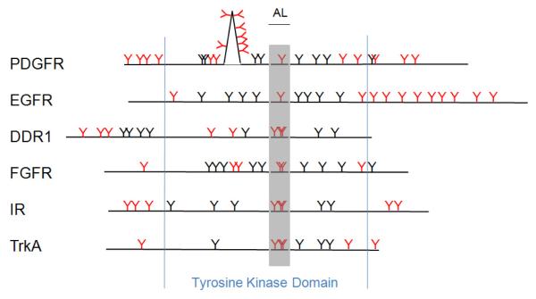
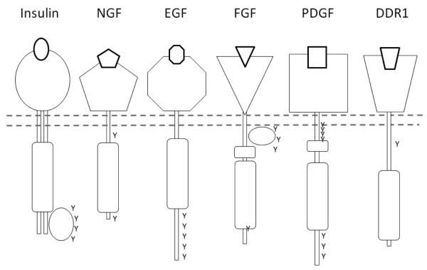
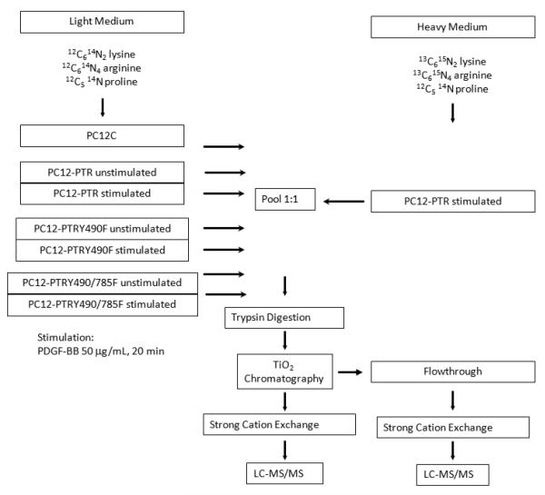
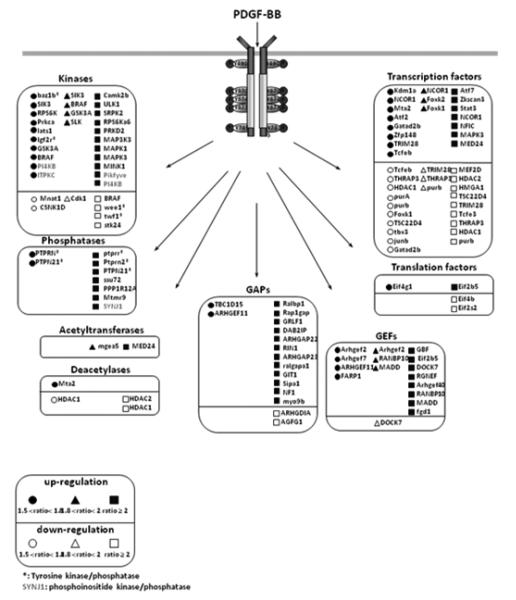
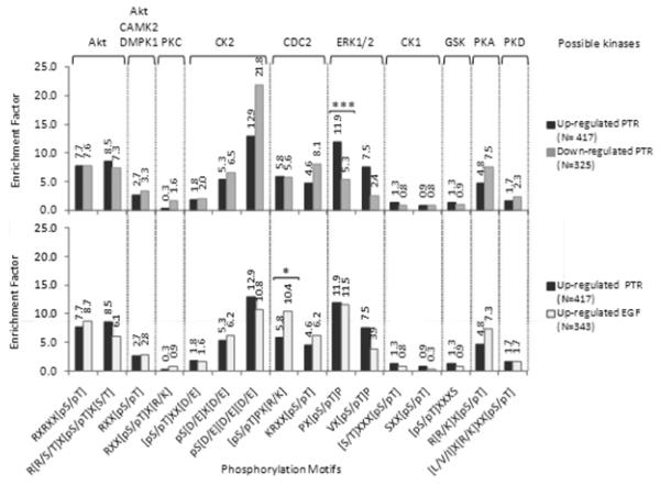
Receptor tyrosine kinases (RTKs) function through protein kinase entities located in the intracellular domain of each protomer. Following activation by ligand binding, they selectively form phosphotyrosine residues by autocatalytic modification. Some of these sites are involved in maintaining the active conformation of the kinase, while others become docking sites for various adaptor/effector/scaffold proteins, which, after complexing with the receptor, then initiate further responses through cascades of post-translational modifications and the generation of lipid second messengers. Although there is substantial overlap in the pathways and activities stimulated by this superfamily, the molecular features of the endodomains of the sub-families and the moieties that they interact with to perpetrate their signals are surprisingly distinct, which may play a significant role in the regulation and responses of the individual RTK types. Some use large scaffold proteins as the basis for most, if not all, of their signal-generating interactions, while others have numerous receptor endodomain phosphotyrosine sites that are quite overlapping in specificity. The members of the Trk family of receptors each have several tyrosine residues that are phosphorylated following stimulation, including those in the kinase activation loop, but there are only two established sites (Y490 and Y785 on TrkA) that are known to be directly involved in signal propagation. Taking advantage of this limited repertoire of docking sites, we have applied phosphoproteomic methods to dissect the signaling responses of both the native protein and derivatives that have had these two sites modified. Interestingly, a clear subset that was not dependent on either docking site was identified. A comparison with a similar set of data for EGFR indicates a considerable degree of similarity in the downstream signaling profile between these two RTKs.
Copyright © 2012 Elsevier Ltd. All rights reserved.
Figures
Similar articles
-
Dissecting the roles of tyrosines 490 and 785 of TrkA protein in the induction of downstream protein phosphorylation using chimeric receptors.J Biol Chem. 2013 Jun 7;288(23):16606-16618. doi: 10.1074/jbc.M113.475285. Epub 2013 Apr 15. J Biol Chem. 2013. PMID: 23589303 Free PMC article.
-
FRS2 proteins recruit intracellular signaling pathways by binding to diverse targets on fibroblast growth factor and nerve growth factor receptors.Mol Cell Biol. 2000 Feb;20(3):979-89. doi: 10.1128/MCB.20.3.979-989.2000. Mol Cell Biol. 2000. PMID: 10629055 Free PMC article.
-
Insulin receptor substrate-1 enhances growth hormone-induced proliferation.Endocrinology. 1999 May;140(5):1972-83. doi: 10.1210/endo.140.5.6724. Endocrinology. 1999. PMID: 10218944
-
The coming of age of phosphoproteomics--from large data sets to inference of protein functions.Mol Cell Proteomics. 2013 Dec;12(12):3453-64. doi: 10.1074/mcp.R113.032862. Epub 2013 Sep 13. Mol Cell Proteomics. 2013. PMID: 24037665 Free PMC article. Review.
-
The FRK/RAK-SHB signaling cascade: a versatile signal-transduction pathway that regulates cell survival, differentiation and proliferation.Curr Mol Med. 2003 Jun;3(4):313-24. doi: 10.2174/1566524033479744. Curr Mol Med. 2003. PMID: 12776987 Review.
Cited by
-
Trk Receptors and Neurotrophin Cross-Interactions: New Perspectives Toward Manipulating Therapeutic Side-Effects.Front Mol Neurosci. 2017 May 3;10:130. doi: 10.3389/fnmol.2017.00130. eCollection 2017. Front Mol Neurosci. 2017. PMID: 28515680 Free PMC article.
-
NGF signaling in PC12 cells: the cooperation of p75(NTR) with TrkA is needed for the activation of both mTORC2 and the PI3K signalling cascade.Biol Open. 2013 Jul 12;2(8):855-66. doi: 10.1242/bio.20135116. eCollection 2013 Aug 15. Biol Open. 2013. PMID: 23951412 Free PMC article.
-
Mechanisms of activation of receptor tyrosine kinases: monomers or dimers.Cells. 2014 Apr 22;3(2):304-30. doi: 10.3390/cells3020304. Cells. 2014. PMID: 24758840 Free PMC article.
-
Role of Epidermal Growth Factor Receptor (EGFR) and Its Ligands in Kidney Inflammation and Damage.Mediators Inflamm. 2018 Dec 23;2018:8739473. doi: 10.1155/2018/8739473. eCollection 2018. Mediators Inflamm. 2018. PMID: 30670929 Free PMC article. Review.
-
EGFRvIII: An Oncogene with Ambiguous Role.J Oncol. 2019 Dec 16;2019:1092587. doi: 10.1155/2019/1092587. eCollection 2019. J Oncol. 2019. PMID: 32089685 Free PMC article. Review.
References
-
- Arkin IT. Structural aspects of oligomerization taking place between the transmembrane alpha-helices of bitopic membrane proteins. Biochim Biophys Acta. 2002;1565(2):347–363. - PubMed
-
- Blume-Jensen p, Hunter T. Oncogenic kinase signaling. Nature. 2001;411:355–365. - PubMed
-
- Bradshaw RA, Dennis EA, editors. Handbook of Cell Signaling. Elsevier Academic Press; San Diego, CA: 2009.
-
- Choudhary C, Mann M. Decoding signalling networks by mass spectrometry-based proteomics. Nat, Revs Mol. Cell Biol. 2010;11:427–439. - PubMed
Publication types
MeSH terms
Substances
Grants and funding
LinkOut - more resources
Full Text Sources
Other Literature Sources
Research Materials
Miscellaneous
