Review
. 2013 May:58:143-52.
doi: 10.1016/j.yjmcc.2012.12.006. Epub 2012 Dec 22.

CaV1.2 signaling complexes in the heart

Affiliations
Review

CaV1.2 signaling complexes in the heart

Robert D Harvey et al. J Mol Cell Cardiol. 2013 May.

Abstract

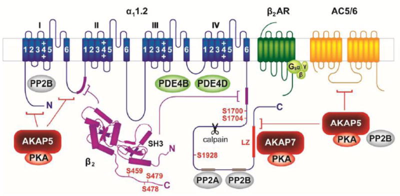
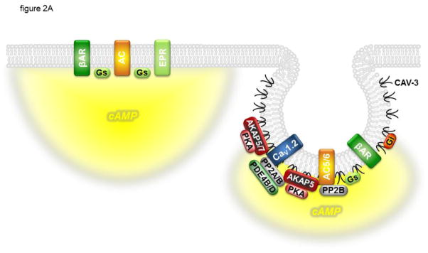
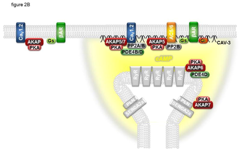
L-type Ca(2+) channels (LTCCs) are essential for generation of the electrical and mechanical properties of cardiac muscle. Furthermore, regulation of LTCC activity plays a central role in mediating the effects of sympathetic stimulation on the heart. The primary mechanism responsible for this regulation involves β-adrenergic receptor (βAR) stimulation of cAMP production and subsequent activation of protein kinase A (PKA). Although it is well established that PKA-dependent phosphorylation regulates LTCC function, there is still much we do not understand. However, it has recently become clear that the interaction of the various signaling proteins involved is not left to completely stochastic events due to random diffusion. The primary LTCC expressed in cardiac muscle, CaV1.2, forms a supramolecular signaling complex that includes the β2AR, G proteins, adenylyl cyclases, phosphodiesterases, PKA, and protein phosphatases. In some cases, the protein interactions with CaV1.2 appear to be direct, in other cases they involve scaffolding proteins such as A kinase anchoring proteins and caveolin-3. Functional evidence also suggests that the targeting of these signaling proteins to specific membrane domains plays a critical role in maintaining the fidelity of receptor mediated LTCC regulation. This information helps explain the phenomenon of compartmentation, whereby different receptors, all linked to the production of a common diffusible second messenger, can vary in their ability to regulate LTCC activity. The purpose of this review is to examine our current understanding of the signaling complexes involved in cardiac LTCC regulation.


Conflict of interest statement

CONFLICT OF INTEREST: THE AUTHORS DECLARE THAT THERE IS NO CONFLICT OF INTEREST

Figures

Figure 1
Figure 1
The CaV1.2 signaling complex includes the α11.2 subunit, β2 subunit, A kinase anchoring proteins 5 and 7 (AKAP5 and AKAP7), protein kinase A (PKA), protein phosphatases 2A and 2B (PP2A and PP2B), the β2-adrenergic receptor (β2AR), adenylyl cyclase 5/6 (AC5/6), stimulatory G protein (Gs), and phosphodiesterases 4B and 4D (PDE4B and PDE4D). PKA phosphorylation sites are indicated in red; sites of interaction with binding partners are indicated by corresponding color-coded segments or brackets. Site cleaved by calpain indicated by scissors. See text for details.
Figure 2
Figure 2
CaV1.2 is regulated by cAMP confined to specific signaling domains. Panel A: β 1- and β2-adrenergic receptors (βARs) associated with CaV1.2 and caveolin-3 (CAV-3) signaling complexes in caveolae can stimulate cAMP production that regulates channel activity. E type prostaglandin receptors (EPRs) and βARs found outside of these signaling complexes can stimulate global changes in cAMP concentration that are not accessible to CaV1.2. Panel B: βARs associated with CaV1.2 and caveolin-3 at dyadic junctions, where caveolae are not present, can stimulate cAMP production that regulates channel activity as well as the activity of the type 2 ryanodine receptor (RyR2) and phospholamban (PLN). βARs not associated with CAV-3 and adenylyl cyclase (AC) are not able to stimulate cAMP production and regulates channel activity. In addition to the CaV1.2 signaling complex described in figure 1, CAV-3 associates with the inhibitory G protein (Gi), and AC5/6 associates with AKAP5 and PP2B. AKAPs also form signaling complexes with PLN and the type 2a sarcoplasmic reticulum Ca2+ ATPase (SERCA2a) as well as RyR2. See text for details.
Figure 2
Figure 2
CaV1.2 is regulated by cAMP confined to specific signaling domains. Panel A: β 1- and β2-adrenergic receptors (βARs) associated with CaV1.2 and caveolin-3 (CAV-3) signaling complexes in caveolae can stimulate cAMP production that regulates channel activity. E type prostaglandin receptors (EPRs) and βARs found outside of these signaling complexes can stimulate global changes in cAMP concentration that are not accessible to CaV1.2. Panel B: βARs associated with CaV1.2 and caveolin-3 at dyadic junctions, where caveolae are not present, can stimulate cAMP production that regulates channel activity as well as the activity of the type 2 ryanodine receptor (RyR2) and phospholamban (PLN). βARs not associated with CAV-3 and adenylyl cyclase (AC) are not able to stimulate cAMP production and regulates channel activity. In addition to the CaV1.2 signaling complex described in figure 1, CAV-3 associates with the inhibitory G protein (Gi), and AC5/6 associates with AKAP5 and PP2B. AKAPs also form signaling complexes with PLN and the type 2a sarcoplasmic reticulum Ca2+ ATPase (SERCA2a) as well as RyR2. See text for details.

References

    1. Dai S, Hall DD, Hell JW. Supramolecular assemblies and localized regulation of voltage-gated ion channels. Physiol Rev. 2009;89:411–52. - PMC - PubMed
    1. Best JM, Kamp TJ. Different subcellular populations of L-type Ca2+ channels exhibit unique regulation and functional roles in cardiomyocytes. J Mol Cell Cardiol. 2012;52:376–87. - PMC - PubMed
    1. Beene DL, Scott JD. A-kinase anchoring proteins take shape. Curr Opin Cell Biol. 2007;19:192–8. - PMC - PubMed
    1. Carafoli E. Calcium signaling: a tale for all seasons. Proc Natl Acad Sci U S A. 2002;99:1115–22. - PMC - PubMed
    1. Clapham DE. Calcium signaling. Cell. 1995;80:259–68. - PubMed

MeSH terms

Substances