

Subretinal drusenoid deposits in non-neovascular age-related macular degeneration: morphology, prevalence, topography, and biogenesis model
- PMID: 23266879
- PMCID: PMC3870202
- DOI: 10.1097/IAE.0b013e31827e25e0
Subretinal drusenoid deposits in non-neovascular age-related macular degeneration: morphology, prevalence, topography, and biogenesis model
Abstract
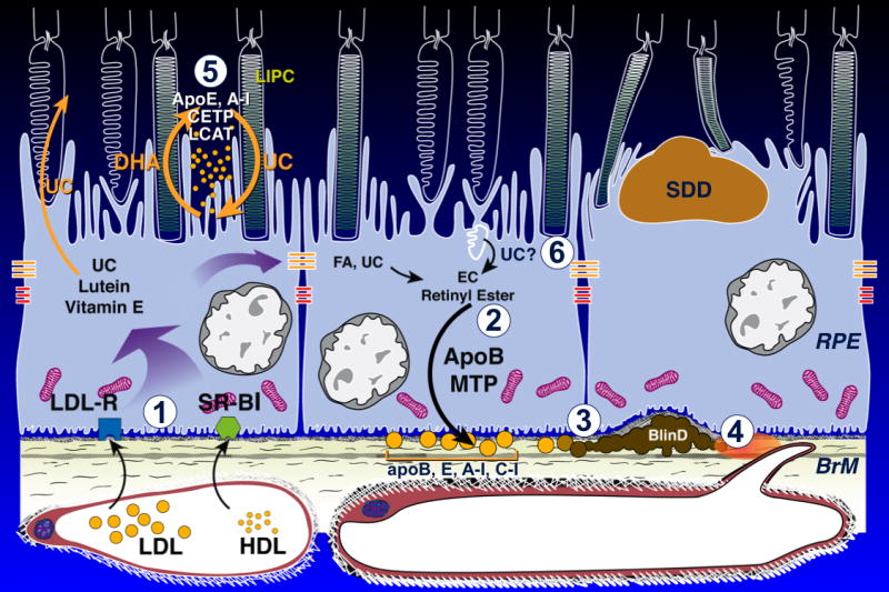
Purpose: To characterize the morphology, prevalence, and topography of subretinal drusenoid deposits, a candidate histological correlate of reticular pseudodrusen, with reference to basal linear deposit (BlinD), a specific lesion of age-related macular degeneration, and to propose a biogenesis model for both lesion.
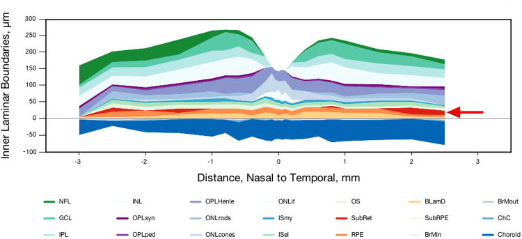
Methods: Donor eyes with median death-to-preservation of 2:40 hours were postfixed in osmium tannic acid paraphenylenediamine and prepared for macula-wide high-resolution digital sections. Annotated thicknesses of 21 chorioretinal layers were determined at standard locations in sections through the fovea and the superior perifovea.
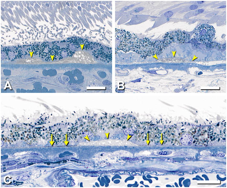
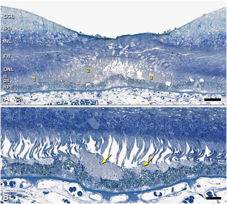
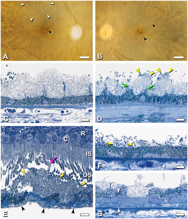
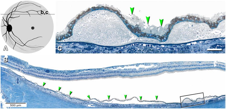
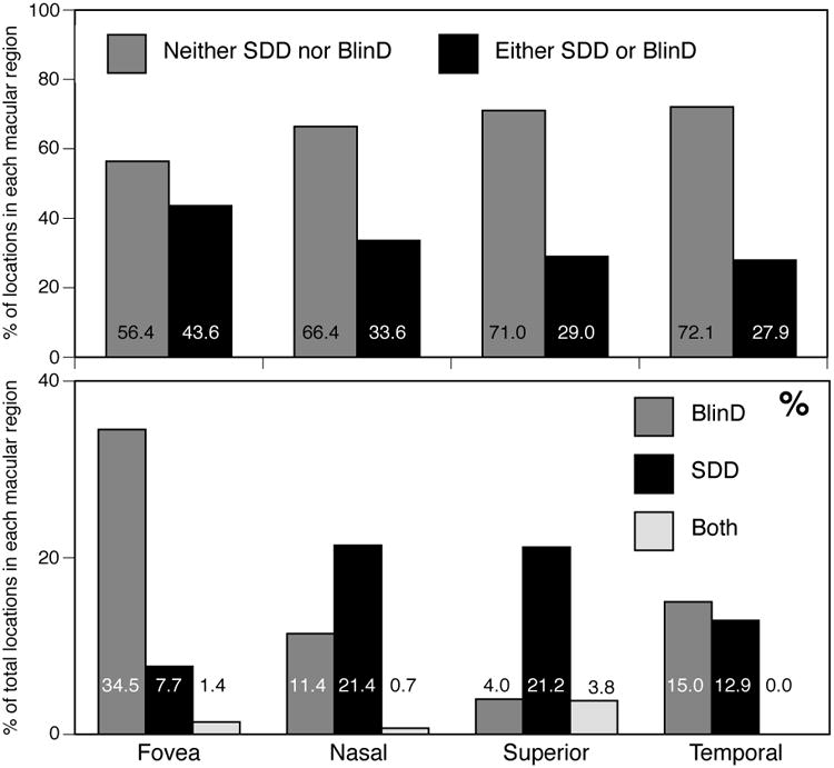
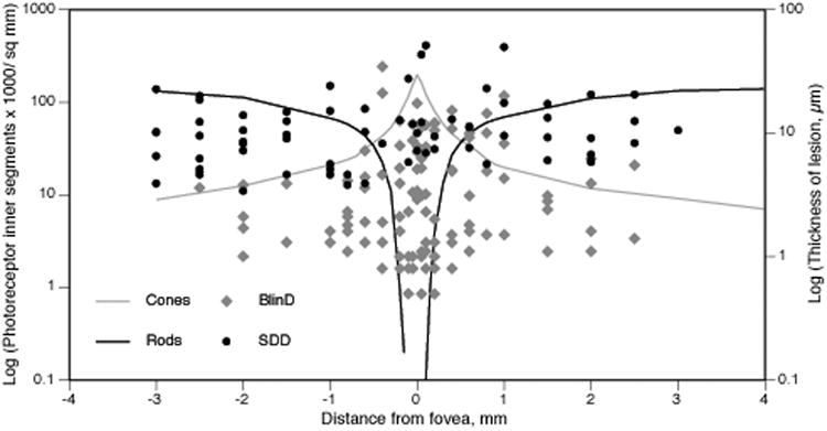
Results: In 22 eyes of 20 white donors (83.1 ± 7.7 years), SDD appeared as isolated or confluent drusenoid dollops punctuated by tufts of retinal pigment epithelium apical processes and associated with photoreceptor perturbation. Subretinal drusenoid deposits and BlinD were detected in 85 and 90% of non-neovascular age-related macular degeneration donors, respectively. Subretinal drusenoid deposit was thick (median, 9.4 μm) and more abundant in the perifovea than in the fovea (P < 0.0001). BlinD was thin (median, 2.1 μm) and more abundant in the fovea than in the perifovea (P < 0.0001).
Conclusion: Subretinal drusenoid deposits and BlinD prevalence in age-related macular degeneration eyes are high. Subretinal drusenoid deposits organized morphology, topography, and impact on surrounding photoreceptors imply specific processes of biogenesis. Contrasting topographies of subretinal drusenoid deposits and BlinD suggest relationships with differentiable aspects of rod and cone physiology, respectively. A 2-lesion 2-compartment biogenesis model incorporating outer retinal lipid homeostasis is presented.
Figures
References
-
- Zweifel SA, Spaide RF, Curcio CA, et al. Reticular pseudodrusen are subretinal drusenoid deposits. Ophthalmology. 2010;117:303–12e.1. - PubMed
-
- Sarks JP, Sarks SH, Killingsworth MC. Evolution of geographic atrophy of the retinal pigment epithelium. Eye. 1988;2:552–577. - PubMed
-
- Sarks S, Cherepanoff S, Killingsworth M, Sarks J. Relationship of basal laminar deposit and membranous debris to the clinical presentation of early age-related macular degeneration. Invest Ophthalmol Vis Sci. 2007;48:968–77. - PubMed
-
- Curcio CA, Presley JB, Medeiros NE, et al. Esterified and unesterified cholesterol in drusen and basal deposits of eyes with age-related maculopathy. Exp Eye Res. 2005;81(6):731–741. - PubMed
Publication types
MeSH terms
Supplementary concepts
Grants and funding
LinkOut - more resources
Full Text Sources
Other Literature Sources
