. 2013 Feb;24(4):440-52.
doi: 10.1091/mbc.E12-10-0749. Epub 2012 Dec 24.

Modular assembly of yeast cytochrome oxidase

Affiliations

Modular assembly of yeast cytochrome oxidase

Gavin P McStay et al. Mol Biol Cell. 2013 Feb.

Abstract

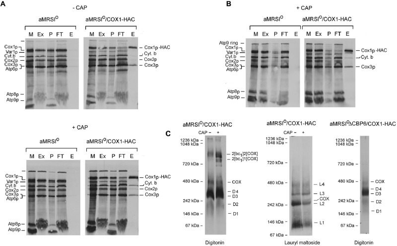
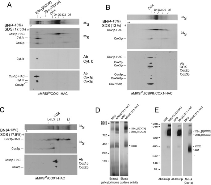
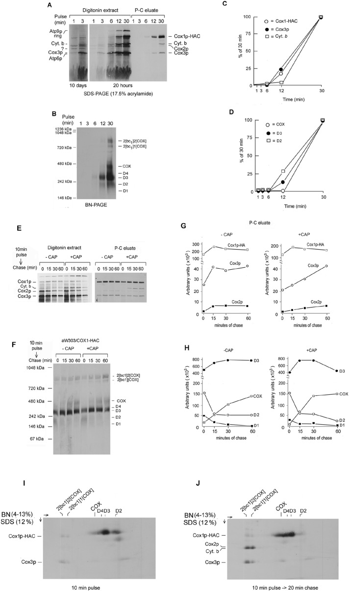
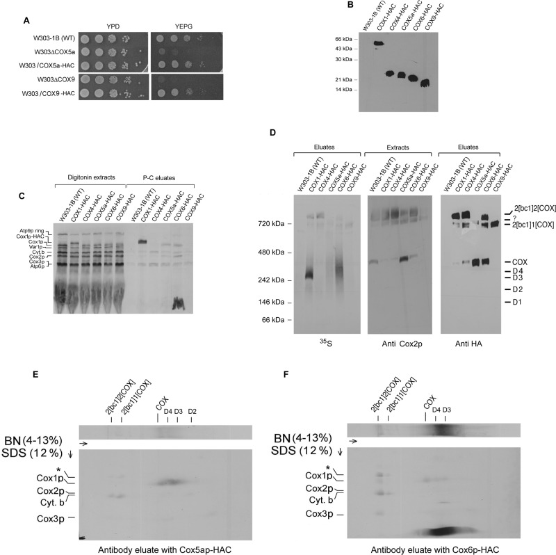
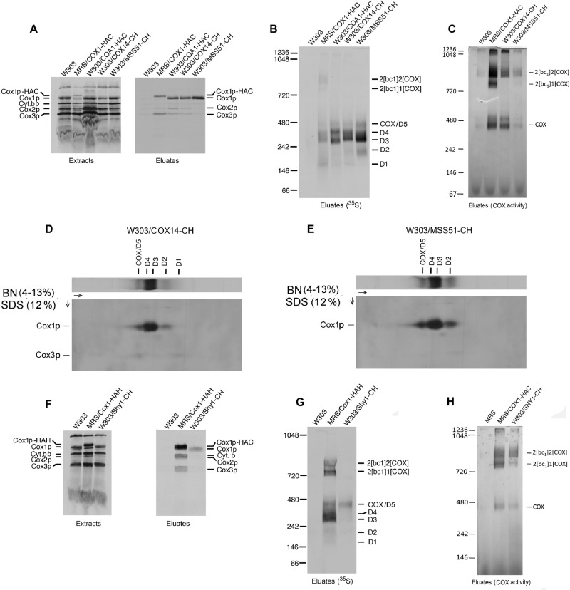
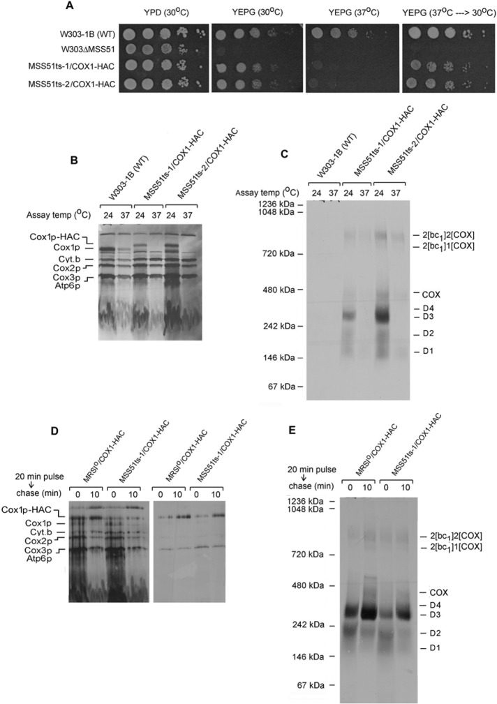
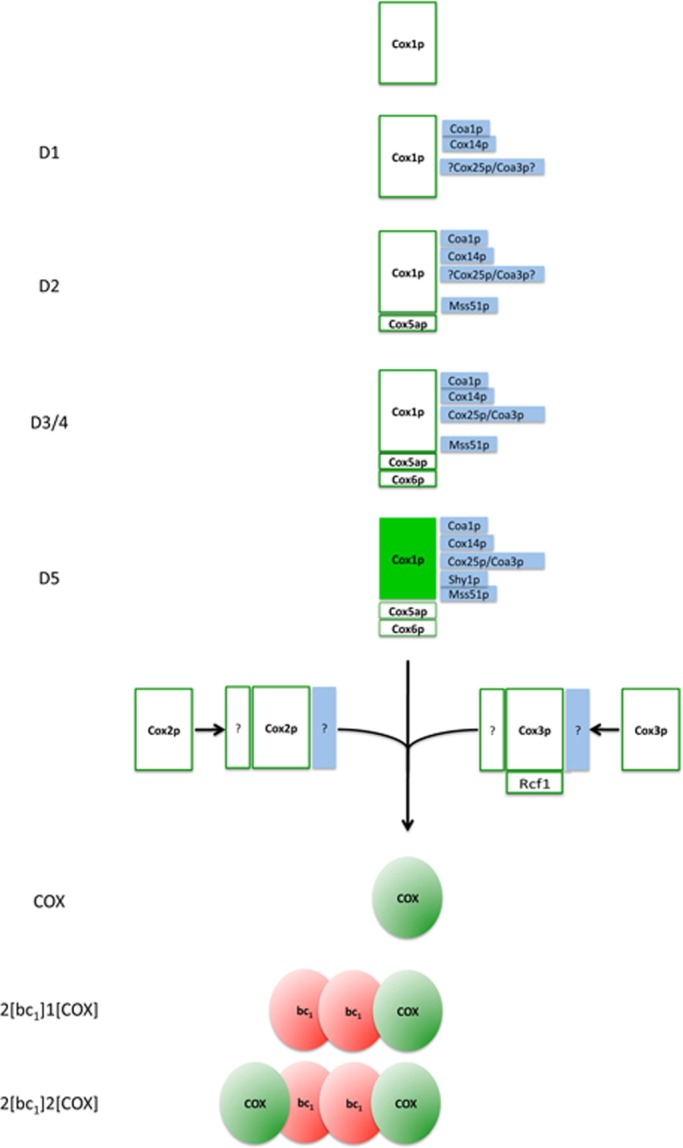
Previous studies of yeast cytochrome oxidase (COX) biogenesis identified Cox1p, one of the three mitochondrially encoded core subunits, in two high-molecular weight complexes combined with regulatory/assembly factors essential for expression of this subunit. In the present study we use pulse-chase labeling experiments in conjunction with isolated mitochondria to identify new Cox1p intermediates and place them in an ordered pathway. Our results indicate that before its assimilation into COX, Cox1p transitions through five intermediates that are differentiated by their compositions of accessory factors and of two of the eight imported subunits. We propose a model of COX biogenesis in which Cox1p and the two other mitochondrial gene products, Cox2p and Cox3p, constitute independent assembly modules, each with its own complement of subunits. Unlike their bacterial counterparts, which are composed only of the individual core subunits, the final sequence in which the mitochondrial modules associate to form the holoenzyme may have been conserved during evolution.


Figures

FIGURE 1:
FIGURE 1:
Analysis of Cox1p complexes in isolated mitochondria. (A) aMRSIo and aMRSIo/COX1-HAC were grown to early stationary phase in YPGal. Cells from one-half of the cultures were used to prepare mitochondria. The other half was inoculated into the starting volumes of fresh YPGal containing 2 mg/ml chloramphenicol, and growth was continued for an additional 2 h before preparation of mitochondria. Mitochondria (500 μg of protein) were labeled at 24°C with a mixture of [35S]methionine and cysteine (Rak et al., 2011). After 20 min, unlabeled methionine and cysteine were added to final concentrations of 1.6 and 0.8 mM, respectively. Mitochondria were centrifuged at 10,000 × gav for 10 min, washed with 0.4 ml of buffer containing 0.6 M sorbitol and 20 mM 4-(2-hydroxyethyl)-1-piperazineethanesulfonic acid (HEPES), pH 7, and suspended at a protein concentration of 10 mg/ml in extraction buffer containing 3% digitonin, 150 mM potassium acetate, 2 mM α-aminocaproic acid, and 20 mM HEPES, pH 7. The suspension was centrifuged at 100,000 × gav for 10 min, and the supernatant (74 μl) was added to 100 μl of packed protein C antibody beads that had been prewashed with wash buffer. The beads were rotated at 4°C for 90 min, centrifuged, and washed three times with 0.5% digitonin in wash buffer. Proteins were eluted by rotation of the washed beads at 4°C for 30 min with 100 μl of elution buffer containing 0.5% digitonin. The following fractions were separated by SDS–PAGE on a 17% polyacrylamide gel: E, eluate from the beads; Ex, digitonin extract; FT, protein fraction that did not adsorb to the antibody beads; M, mitochondria; P, pellet after digitonin extraction. All of the fractions loaded on the gel were adjusted to the starting volume of mitochondria (equivalent to 20 μg of protein). (B) aMRSIo and aMRSIo/COX1 were grown as in A with a 2-h incubation in medium containing chloramphenicol. The mitochondria were labeled and fractionated as in A, except that the extraction buffer contained 1% lauryl maltoside instead of digitonin and the antibody beads were washed and eluted in the presence of 0.2% lauryl maltoside. (C) Eluates of the samples obtained from mitochondria grown in the presence and absence of chloramphenicol were mixed with 0.14 volume of 7× sample buffer (Wittig et al., 2006) and separated by BN-PAGE. The sample shown in the extreme right was obtained from a cbp6 mutant (aMRSIoΔCBP6/COX1-HAC) grown in the absence of chloramphenicol.
FIGURE 2:
FIGURE 2:
Two-dimensional BN- and SDS–PAGE analysis of radiolabeled Cox1p complexes. (A) Mitochondria were prepared from aMRSIo/COX1-HAC grown in YPGal to early stationary phase and transferred for 2 h to fresh YPGal medium containing 2 mg/ml chloramphenicol. Mitochondria labeled with [35S]methionine plus cysteine for 20 min were extracted with digitonin and the extracts purified on protein C antibody beads as in Figure 1A. Proteins were separated by BN-PAGE in the first dimension. A strip of the first dimension gel was soaked for 30 min in a buffer containing 0.1% SDS and 100 mM Tris-Cl, pH 6.8, and was layered on top of a 17% SDS-polyacrylamide gel for separation in the second dimension (Laemmli, 1970). Proteins were transferred to a polyvinylidene fluoride (PVDF) membrane and exposed to x-ray film. The radiolabeled band migrating midway between Cox1p and Cox3p in the region corresponding to the supercomplexes was confirmed to be cytochrome b by Western analysis (bottom). (B) Same as A, except that mitochondria were obtained from the cbp6 mutant aMRSIoΔCBP6/COX1-HAC grown in the absence of chloramphenicol. After autoradiography the gel was treated sequentially with 1) a polyclonal antibody against yeast cytochrome oxidase, 2) a monoclonal antibody against Cox2p, and 3) a monoclonal antibody against Cox3p. The COX antibody detects Cox1p and Cox4-9. (C) Same as A, except that the labeled mitochondria were extracted with lauryl maltoside as in Figure 1B. (D) Digitonin extracts of mitochondria from MRS-3B and MRS/COX1-HAC were fractionated on protein C antibody beads as in Figure 1A. Extracts and eluates from 50 and 170 μg of starting mitochondria protein were separated by BN-PAGE. The gel was immersed in a solution containing 0.5 mg/ml DAB and 1 mg/ml horse heart cytochrome c for several hours. The reaction was stopped with 45% methanol/5% acetic acid. Molecular weight markers were visualized by Coomassie R-250 staining after documentation of the in-gel activity. (E) Digitonin extracts of mitochondria from MRS-3B and MRS/COX1-HAC were solubilized in 3% digitonin and fractionated on protein C antibody beads. Eluates equivalent to 450 μg of starting mitochondrial protein were separated by BN-PAGE, transferred to a PVDF membrane, and reacted with antibodies against Cox2p and Cox3p. The Cox3p immunoblot was stripped and reprobed with anti-HA antibody to detect Cox1p-HAC.
FIGURE 3:
FIGURE 3:
Kinetics of Cox1p-HAC incorporation into COX subcomplexes. (A) aMRSIo/COX1-HAC mitochondria were labeled with [35S]methionine plus cysteine for the indicated times and immediately added to an equal volume of 4% digitonin in extraction buffer and centrifuged. The digitonin extracts were further purified on protein C antibody beads as in Figure 1A. Samples of the extracts and eluates from the beads were separated by SDS–PAGE on a 17% polyacrylamide gel, transferred to nitrocellulose, and exposed to x-ray film for 20 h and for 10 d. (B) The eluates from the antibody beads were separated by BN-PAGE, transferred to PVDF membrane, and exposed to x-ray film. (C) Cox1p-HAC, Cox3p, and cytochrome b eluted from the antibody beads were quantified with a PhosphorImager. The results are plotted as percentage of the values obtained at 30 min. (D) The bands corresponding to COX, D2, and D3 were quantified as in C. (E) aMRSIo/COX1-HAC was grown to late–log phase in YPGal. Cells were harvested and equal parts inoculated into fresh YPGal without (–CAP) and with 2 mg/ml chloramphenicol (+CAP). The cultures were grown for an additional 2.5 h before preparation of mitochondria. Mitochondria equivalent to 250 μg of protein in a final volume of 80 μl were labeled for 10 min at 24°C with [35S]methionine plus cysteine. Translation was terminated by addition of puromycin to a final concentration of 100 μM, and a sample (0 time) representing 25% of the total translation mix was added to 1.2 volumes of 4% digitonin in extraction buffer and centrifuged at 100,000 × gav for 10 min. Identical-size samples were extracted with digitonin after 15, 30, and 60 min of incubation at 24°C. Digitonin extracts and eluates from the protein C antibody beads, representing 5 and 25% of the starting material, respectively, were separated by SDS–PAGE and radiolabeled proteins visualized as in A. (F) The remainder of the eluates from the antibody beads were separated by BN- PAGE and radiolabeled proteins visualized as in B. (G) The radiolabeled bands corresponding the Cox1p-HAC, Cox2, and Cox3 in the protein C eluates were quantified as in C. (H) The bands identified as COX and the three subcomplexes D2, D3, and D4 of the native gel shown in Figure 3F were quantified with a PhosphorImager as in D. (I, J) Mitochondria from aMRSIo/COX1-HAC were labeled at 24°C with [35S]methionine and cysteine for 10 min. Translation was terminated by addition of puromycin. After incubation for an additional 20 min mitochondria were extracted with digitonin as in E, and Cox1p-HAC was purified on the antibody beads. The eluates from the beads were separated by BN-PAGE on a 3–14% polyacrylamide gel in the first dimension and by SDS–PAGE on a 17% gel in the second dimension. Proteins were transferred to PVDF and visualized by exposure to x-ray film.
FIGURE 4:
FIGURE 4:
Subunit composition of Cox1p intermediates. (A) Serial dilutions of the wild-type W303-1B, a cox5a (W303ΔCOX5a)- and a cox9 (W303ΔCOX9)-null mutant, and null mutants expressing HAC-tagged Cox5ap and Cox9p were serially diluted and spotted on rich glucose (YPD) and rich glycerol plus ethanol (YEPG) plates. The plates were photographed after 2 d at 30°C. (B) Mitochondria (12.5 μg of protein) from W303-1B and strains of yeast expressing Cox4p, Cox5ap, Cox6p, and Cox9p with C-terminal HAC double tags were separated by SDS–PAGE on a 12% polyacrylamide gel. Proteins were transferred to nitrocellulose and probed with a mouse monoclonal antibody against the HA epitope as in Figure 2A. (C) Mitochondria were labeled for 20 min at room temperature with [35S]methionine and cysteine. Translation was terminated with puromycin, and incubation continued for an additional 10 min. The labeled mitochondria were extracted by addition of 1.2 volumes of 4% digitonin and tagged subunits purified on protein C antibody beads as in Figure 1A. Samples of extracts and eluates were separated by SDS–PAGE on a 17% polyacrylamide gel. (D) The digitonin extracts and eluates obtained in C were separated by BN-PAGE. After transfer to PVDF, the blot with the eluates was first exposed to x-ray film and then probed with a mouse monoclonal antibody against the HA epitope. The blot with the extracts was immunostained with a monoclonal antibody against Cox2p. (E, F) The eluates with Cox5ap-HAC and Cox6p-HAC were separated by BN-PAGE in the first dimension and by SDS–PAGE in the second dimension. Proteins were transferred to nitrocellulose and exposed to x-ray film.
FIGURE 5:
FIGURE 5:
Analysis of Cox14p, Mss51p, Coa1p, and Shy1p in the Cox1p intermediates. W303-1B, MRS/COX1-HAC, W303/COA1-HAC, W303/COX14-CH, and W303/MSS51-CH were grown in YPGal and mitochondria labeled with [35S]methionine plus cysteine. Puromycin was added after 30 min, and incubation continued for an additional 10 min. Mitochondria (450 μg of protein) were extracted with digitonin and the tagged proteins purified on protein C antibody beads as in Figure 1A. Radiolabeled mitochondrial gene products were visualized after separation of the digitonin extracts and eluates on a 17% polyacrylamide gel by SDS–PAGE (A) and BN-PAGE (B). (C) The eluates from the antibody beads were separated by BN-PAGE and stained for COX activity as in Figure 1D. (D, E) Mitochondria were labeled and extracted with digitonin, and the extracts were purified on antibody beads as in A. Eluates from the beads were separated by 2D gel electrophoresis. Proteins were transferred to a PVDF membrane and exposed to x-ray film. (F) Mitochondria from W303-1B, MRS/COX1-HAH, and W303/SHY1-CH were labeled and extracted as in A. The digitonin extracts were incubated with Ni-NTA beads (Qiagen, Valencia, CA) for 2 h in the presence of 400 mM NaCl, 20 mM imidazole, and 0.5% digitonin, pH 8.0. The resin was washed three times in the same buffer and proteins eluted with 150 mM imidazole, pH 7.4, in 0.5% digitonin. Samples of the extracts and eluates were separated by SDS–PAGE on a 17.5% polyacrylamide gel, transferred to nitrocellulose, and exposed to x-ray film. (G) The eluates from Ni-NTA were separated by BN-PAGE. Proteins were transferred to PVDF membrane and exposed to x-ray film. (H) Mitochondria from W303-1B, MRS/COX1-HAH, and W303/SHY1-CH were extracted as in C. The digitonin extracts were incubated with Ni-NTA resin and eluted as in F and stained for COX activity as in C.
FIGURE 6:
FIGURE 6:
Cox1p intermediates in an mss51 ts mutant. (A) Serial dilution of the wild-type W303-1B, the mss51-null mutant (W303ΔMSS51), and two independent mss51 ts mutants, MSS51ts-1/COX1-HAC and MSS51ts-2/COX1-HAC, were spotted on YPD and YEPG. The plates were incubated for 2 d at the indicated temperatures. The YEPG plate that had been incubated at 37°C was transferred to 30°C and incubated for an additional 2 d (far right). (B, C) Mitochondria were prepared from the wild-type strain W303-1B and the two mss51 ts mutants grown at 30°C. They were preincubated either at 24 or 37°C for 5 min before addition of [35S]methionine and cysteine and further incubated at the same temperatures for 30 min. Mitochondria were extracted with 1.2 volumes of 4% digitonin and the extracts purified on protein C antibody beads as in Figure 1A. The extracts were separated by SDS–PAGE on a 17% polyacrylamide gel (B) and the eluates from the beads by BN-PAGE (C). Proteins were transferred either to nitrocellulose or a PVDF membrane and exposed to x-ray film. (D) Mitochondria from the wild-type strain MRSIo/COX1-HAC and from the mss51 ts mutant were labeled for 20 min at 24°C with [35S]methionine and cysteine. Puromycin was added, and incubation continued for another 10 min at 37°C before extraction with digitonin. The extracts were purified on antibody beads, and samples of extract and eluates were separated by SDS–PAGE on a 17% polyacrylamide gel. Proteins were transferred to nitrocellulose and exposed to x-ray film. (E) The eluates from D were separated on a blue native gel, transferred to a PVDF membrane, and exposed to x-ray film.
FIGURE 7:
FIGURE 7:
Scheme of cytochrome oxidase assembly. Newly translated Cox1p interacts with Cox14p and Coa1p to form complex D1. Cox25p/Coa3p was detected in D2, D3, D4, and COX/D5 (unpublished data). Owing to poor labeling of D1, the association of Cox25p/Coa3p with this intermediate is uncertain but is provisionally indicated, mainly because of the similarity of its behavior to that of Cox14p. Mss51p and subunit Cox5ap associate with D1 to form complex D2, which is converted to D3 by addition of Cox6p. D4 migrates separately from complex D3, but the compositional difference between the two is not known. D5, which migrates at the position of COX in the native gel, contains Shy1p in addition to the aforementioned components. D5 is proposed to be the last Cox1p intermediate before it combines with Cox2p and Cox3p, the other two mitochondrially translated subunits that make up the core of the enzyme. The completion of COX biogenesis is followed by its interaction with dimeric bc1 complex. Ssc1p (Fontanesi et al., 2010, 2011) and Coa2 (Pierrel et al., 2008; Bestwick et al., 2010), which have also been implicated in the Cox1p biogenesis pathway, have not been examined and are not shown in this scheme.

Similar articles

Cited by

References

    1. Barrientos A, Zambrano A, Tzagoloff A. Mss51p and Cox14p jointly regulate mitochondrial Cox1p expression in Saccharomyces cerevisiae. EMBO J. 2004;23:3472–3482. - PMC - PubMed
    1. Bestwick MM, Jeong Y, Khalimonchuk O, Kim Y, Winge DR. Analysis of Leigh syndrome mutations in the yeast SURF1 homolog reveals a new member of the cytochrome oxidase assembly factor family. Mol Cell Biol. 2010;30:4480–4491. - PMC - PubMed
    1. Bonnefoy N, Fox TD. Directed alteration of Saccharomyces cerevisiae mitochondrial DNA by biolistic transformation and homologous recombination. Methods Mol Biol. 2007;372:153–166. - PMC - PubMed
    1. Carr HS, Winge DR. Assembly of cytochrome c oxidase within the mitochondrion. Acc Chem Res. 2003;36:309–316. - PubMed
    1. Choquet Y, Wostrikoff K, Rimbault B, Zito F, Girard-Bascou J, Drapier D, Wollman FA. Assembly-controlled regulation of chloroplast gene translation. Biochem Soc Trans. 2001;29:421–426. - PubMed

Publication types

MeSH terms