. 2013 Jan 14;210(1):23-30.
doi: 10.1084/jem.20121072. Epub 2012 Dec 24.

The intramembrane protease Sppl2a is required for B cell and DC development and survival via cleavage of the invariant chain

Affiliations

The intramembrane protease Sppl2a is required for B cell and DC development and survival via cleavage of the invariant chain

Daniel R Beisner et al. J Exp Med. .

Abstract

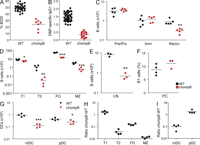
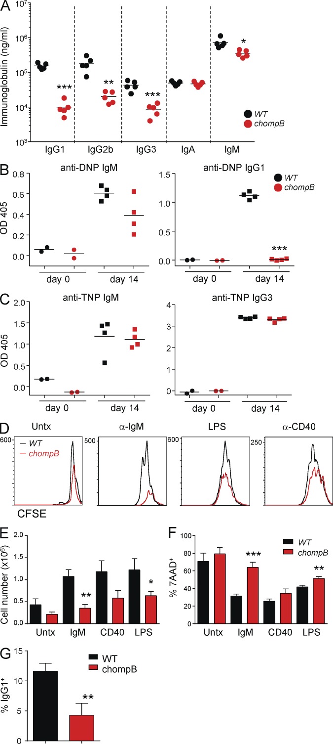
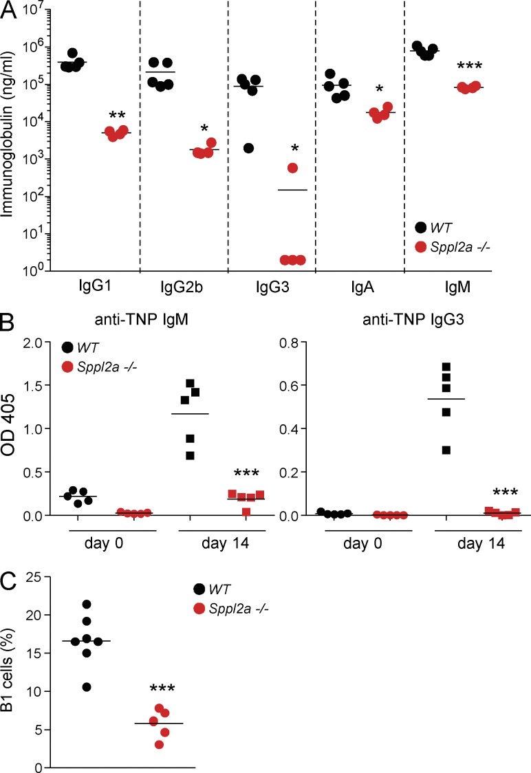
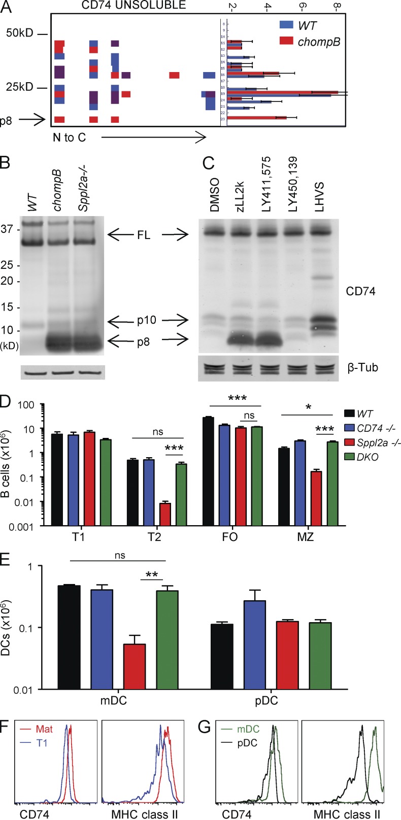
B cell development requires tight regulation to allow for the generation of a diverse repertoire while preventing the development of autoreactive cells. We report, using N-ethyl-N-nitrosourea (ENU)-induced mutagenesis, the identification of a mutant mouse (chompB) with a block in early B cell development. The blockade occurs after the transitional 1 (T1) stage and leads to a decrease in mature B cell subsets and deficits in T cell-dependent antibody responses. Additionally, chompB mice have decreases in myeloid dendritic cells (DCs). The mutation was mapped to the intramembrane protease signal peptide peptidase-like 2a (Sppl2a), a gene not previously implicated in immune cell development. Proteomic analysis identified the invariant chain (CD74) as a key substrate of Sppl2a and suggests that regulated intramembrane proteolysis of CD74 by Sppl2a contributes to B cell and DC survival. Moreover, these data suggest that modulation of Sppl2a may be a useful therapeutic strategy for treatment of B cell dependent autoimmune disorders.


Figures

Figure 1.
Figure 1.
chompB mice have a block in B cell and DC development. (A and B) Mutagenized mice were screened for blood B cells by B220 staining (A) and DNP-KLH–specific IgG by ELISA (B). (C) BM B cells were enumerated by flow cytometry using the following criteria: pre/pro, B220+IgM; immature, B220+IgM+CD24+; mature/recirculating, B220+IgM+CD24. (D) Splenic B cell subsets were enumerated using the following criteria: T1, IgMhiCD21; T2, IgMhiCD21hiCD23+; FO, IgM+CD21lo; MZ, IgMhiCD21hiCD23. (E) B cell numbers in the lymph nodes were determined by gating on B220+ cells. (F) B1 cell numbers from the peritoneal cavity were enumerated using CD19+B220intCD5int. (G) DC numbers were determined using the following criteria: mDC, CD19CD3NK1.1CD11chi; pDC, CD19CD3NK1.1CD11c+PDCA1+. (H and I) 50/50 Mixed BM chimeras were analyzed by flow cytometry 8–12 wk after reconstitution. Data are expressed as the ratio of chompB to WT cells. Analysis of the B cells (H) and DCs (I) was performed using the gating schemes as in D and E after gating on CD45 variants to determine donor. *, P < 0.05; **, P < 0.01; ***, P < 0.001 (mean, n = 3–5 in at least two independent experiments).
Figure 2.
Figure 2.
Defective T cell–dependent antibody responses and B cell activation in chompB mutants. (A) Ig levels were analyzed in 6–12-wk-old WT and chompB mutant mice. (B) 6–12-wk-old WT and chompB mice were immunized with DNP-KLH and, after 14 d, DNP-specific antibody levels were determined by ELISA. (C) 6–12-wk-old WT and chompB mice were immunized with TNP-Ficoll and, after 14 d, TNP-specific antibody levels were determined by ELISA. (D–F) Purified B cells from WT and chompB mice were labeled with CFSE and cultured with the indicated stimuli along with 10 ng/ml rIL-4 for 72 h. CFSE dilution and cell death were then assessed by flow cytometry, and total cell numbers were determined by cell counting. (G) Expression of surface IgG1 in response to anti-CD40 and IL-4 was assessed by flow cytometry after 96 h of culture (mean, n = 4–5 from two independent experiments, A–C; results are representative of at least three replicates from two independent experiments, D; mean and SEM of four independent experiments, E and F; and mean and SEM, n = 4 from two independent experiments, G). *, P < 0.05; **, P < 0.01; ***, P < 0.001.
Figure 3.
Figure 3.
Mutation in Sppl2a is responsible for the observed defects in chompB mice. (A and B) HEK293T cells were cotransfected with the indicated plasmids, and TNF and Sppl2a levels were determined by Western blot. TNF-NTF (N-terminal fragment) was first normalized to full length TNF, followed by normalization to samples without Sppl2a for quantification (representative of four independent experiments for A and mean, n = 4 from four independent experiments for B). (C) U2OS cells were cotransfected with V5-mSppl2a together with Rab5-RFP. Cells were then stained with an anti-V5 antibody and Hoechst dye and analyzed by microscopy (representative of two independent experiments). Bars, 20 µm. (D and E) Splenocytes from WT and Sppl2a−/− mice were analyzed by flow cytometry as described in Fig. 1 (mean, n = 5 from three independent experiments). (F) 6–12-wk-old WT and Sppl2a−/− mice were immunized with DNP-KLH. DNP-specific antibody levels were determined by ELISA after 14 d (mean, n = 4–5 from two independent experiments). ***, P < 0.001.
Figure 4.
Figure 4.
The chompB mutation in Sppl2a is a hypomorphic allele. (A) Plasma Ig levels were determined from WT and Sppl2a−/− mice (mean, n = 4–5 from two independent experiments). (B) 6–12-wk-old WT and Sppl2a−/− mice were immunized with TNP-Ficoll and TNP-specific antibody levels were determined by ELISA 14 d later (mean, n = 4–5, two independent experiments). (C) B1 cell numbers from the peritoneal cavity were defined as in Fig. 1 (mean, n = 5–7 from three independent experiments). *, P < 0.05; **, P < 0.01; ***, P < 0.001.
Figure 5.
Figure 5.
Sppl2a-mediated cleavage of CD74 is responsible for the B cell developmental blockade in chompB mice. (A) Peptograph of CD74 from WT and chompB membrane-enriched fractions. The left side depicts the weight in kD and the right side depicts the frequency. (B) Western blot analysis of sorted mature B cells (CD24loCD21int) using antibodies to CD74 and β-tubulin (representative of two independent experiments). (C) CD74 cleavage in A20 cells assessed by Western blot (results are representative of at least three independent experiments). (D and E) B cell and DC numbers were determined using the same parameters in Fig. 1 (mean, n = 4 from three independent experiments). (F and G) Intracellular CD74 and MHC class II levels were determined using the gating schemes in Fig. 1 for the indicated B cell (F) and DC (G) populations (n = 3, from two independent experiments). *, P < 0.05; **, P < 0.01; ***, P < 0.001.

Similar articles

Cited by

References

    1. Allman D., Pillai S. 2008. Peripheral B cell subsets. Curr. Opin. Immunol. 20:149–157 10.1016/j.coi.2008.03.014 - DOI - PMC - PubMed
    1. Becker-Herman S., Arie G., Medvedovsky H., Kerem A., Shachar I. 2005. CD74 is a member of the regulated intramembrane proteolysis-processed protein family. Mol. Biol. Cell. 16:5061–5069 10.1091/mbc.E05-04-0327 - DOI - PMC - PubMed
    1. Beisner D.R., Chu I.H., Arechiga A.F., Hedrick S.M., Walsh C.M. 2003. The requirements for Fas-associated death domain signaling in mature T cell activation and survival. J. Immunol. 171:247–256 - PubMed
    1. Bergmann H., Yabas M., Short A., Miosge L., Barthel N., Teh C.E., Roots C.M., Bull K.R., Jeelall Y., Horikawa K., et al. 2013. B cell survival, surface BCR and BAFFR expression, CD74 metabolism, and CD8− dendritic cells require the intramembrane endopeptidase SPPL2A. J. Exp. Med. 210:31–40 - PMC - PubMed
    1. Beutler B., Jiang Z., Georgel P., Crozat K., Croker B., Rutschmann S., Du X., Hoebe K. 2006. Genetic analysis of host resistance: Toll-like receptor signaling and immunity at large. Annu. Rev. Immunol. 24:353–389 10.1146/annurev.immunol.24.021605.090552 - DOI - PubMed

MeSH terms