. 2013 Jan 14;210(1):31-40.
doi: 10.1084/jem.20121076. Epub 2012 Dec 24.

B cell survival, surface BCR and BAFFR expression, CD74 metabolism, and CD8- dendritic cells require the intramembrane endopeptidase SPPL2A

Affiliations

B cell survival, surface BCR and BAFFR expression, CD74 metabolism, and CD8- dendritic cells require the intramembrane endopeptidase SPPL2A

Hannes Bergmann et al. J Exp Med. .

Abstract

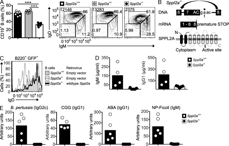
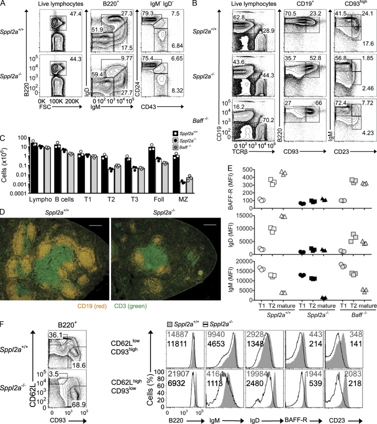
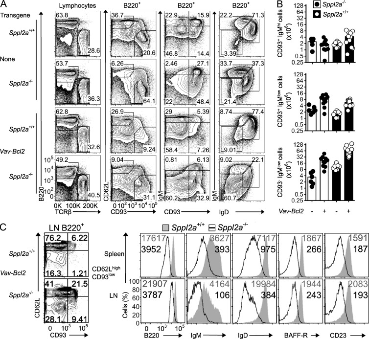
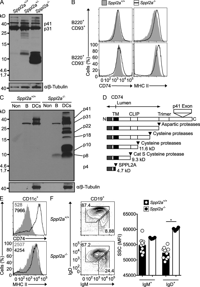
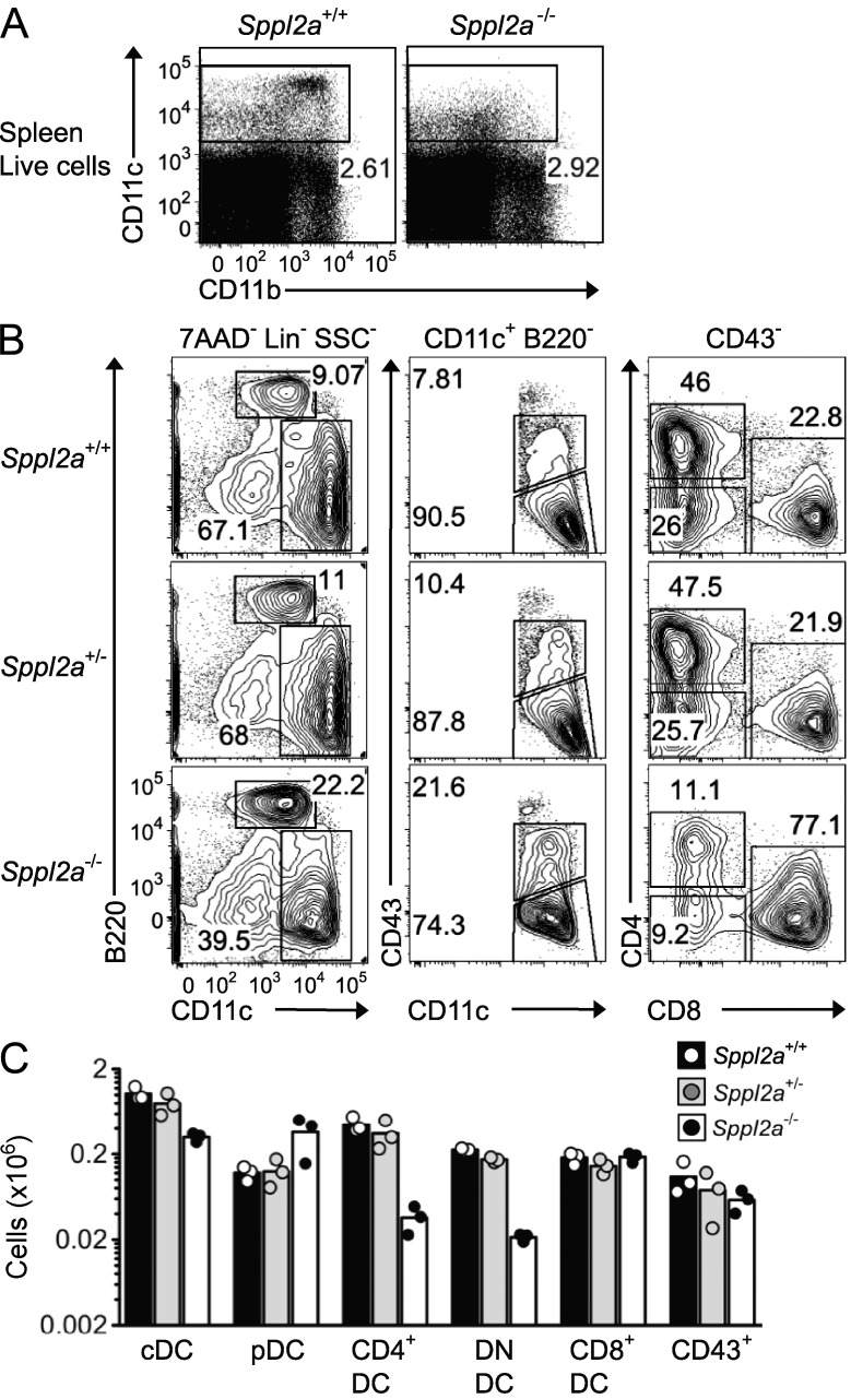
Druggable proteins required for B lymphocyte survival and immune responses are an emerging source of new treatments for autoimmunity and lymphoid malignancy. In this study, we show that mice with an inactivating mutation in the intramembrane protease signal peptide peptidase-like 2A (SPPL2A) unexpectedly exhibit profound humoral immunodeficiency and lack mature B cell subsets, mirroring deficiency of the cytokine B cell-activating factor (BAFF). Accumulation of Sppl2a-deficient B cells was rescued by overexpression of the BAFF-induced survival protein B cell lymphoma 2 (BCL2) but not BAFF and was distinguished by low surface BAFF receptor and IgM and IgD B cell receptors. CD8-negative dendritic cells were also greatly decreased. SPPL2A deficiency blocked the proteolytic processing of CD74 MHC II invariant chain in both cell types, causing dramatic build-up of the p8 product of Cathepsin S and interfering with earlier steps in CD74 endosomal retention and processing. The findings illuminate an important role for the final step in the CD74-MHC II pathway and a new target for protease inhibitor treatment of B cell diseases.


Figures

Figure 1.
Figure 1.
Humoral immunodeficiency in Sppl2a−/− mice. (A) Percentage of CD19+ B cells among blood lymphocytes and representative IgM/IgD flow cytometric plots gated on CD19+ B cells from mice of the indicated genotypes. Numbers in top left corner are geometric mean fluorescence intensity of IgD, and other numbers are percentage of cells in each gate. ****, P < 0.0001. (B) Schematic of Sppl2a splice-donor mutation, the resulting skipping of exon 7, and the effect on the SPPL2A protein. (C) Surface IgD on GFP+IgM+ Sppl2a−/− B cells from IL-7 bone marrow cultures transduced with a retroviral vector encoding wild-type SPPL2A and GFP (thick line) or empty vector encoding GFP alone (thin line), compared with Sppl2a+/+ B cells transduced with empty GFP vector (shaded gray). (D) ELISA analysis of total serum IgM and IgG1 in unimmunized animals. (E) ELISA analysis of serum antibodies to B. pertussis and CGG 2 wk after immunization, against the ABA 4 wk after immunization, and against NP-Ficoll 6 d after booster immunization. Bars represent the mean, and each symbol represents a single mouse. Data are representative of more than five experiments with at least three animals per group in each (A), one experiment (B and C) or two (D), and one experiment with at least five to seven mice per group (E).
Figure 2.
Figure 2.
Mature Sppl2a−/− B cells fail to up-regulate BAFFR or IgD and do not accumulate in the periphery. (A) Representative flow cytometric plots and percentages of bone marrow B cell subsets from homozygous mutant and wild-type mice. (B) Representative flow cytometric plots and percentages of spleen B cell subsets from Sppl2a−/−, Baff−/−, and wild-type (Sppl2a+/+) mice. (C) Number of cells of the indicated subsets in spleens from Sppl2a−/−, Baff−/−, and Sppl2a+/+ mice: Lympho, lymphocytes; Foll, follicular B cells; MZ, marginal zone B cells. Bars show mean, and each symbol is a single mouse. (D) Spleen cryosections from Sppl2a+/+ and Sppl2a−/− mice were stained with fluorescently labeled antibodies to detect B cells (CD19) and T cells (CD3). Bars, 200 µm. (E) Mean fluorescence intensity (MFI) of staining for BAFFR, IgD, and IgM on the indicated B cell subsets in spleens from Sppl2a−/−, Baff−/−, and Sppl2a+/+ mice. (F) Representative flow cytometric staining presented as overlay histograms for B220, IgM, IgD, BAFFR, and CD23 on Sppl2a+/+ (shaded gray) and Sppl2a−/− (black line) immature (CD62LlowCD93high) or mature (CD62LhighCD93low) splenic B cells. The numbers show the MFI for Sppl2a+/+ (gray; top) and Sppl2a−/− cells (black; bottom). Data are representative of at least three experiments with three mice (A–C and E), two experiments with one to three mice per group (D), or one experiment with three mice per group (F). Pregates for FACS plots are indicated above each column.
Figure 3.
Figure 3.
Overexpression of antiapoptotic BCL2 in Sppl2a−/− mice rescues accumulation of mature B cells. (A) Representative flow cytometric plots of lymphocytes in the spleen from mice of the indicated Sppl2a and Vav-Bcl2 genotypes. Pregates for plots are indicated above the columns. (B) Number of B cells of the indicated subsets in spleens from Sppl2a+/+ and Sppl2a−/− mice, either transgenic for Vav-Bcl2 or not. Bars show mean, and each symbol is a single mouse. (C) Representative flow cytometric plots of CD62L and CD93 expression on B cells from subcutaneous LNs in Vav-Bcl2 transgenic mice of the indicated genotype. Overlay of flow cytometric histograms for B220, IgM, IgD, BAFFR, and CD23 cell surface expression on CD62LhighCD93low mature B cells in spleen (top) or LNs (bottom) from Sppl2a+/+ (shaded gray) and Sppl2a−/− (black line) mice. Numbers in the plots indicate geometric mean fluorescence intensity for Sppl2a+/+ (gray; top) and Sppl2a−/− cells (black; bottom). Data are representative of one to three experiments with three to four mice per group (A and C) or combined from four experiments (B).
Figure 4.
Figure 4.
Proteolytic processing and intracellular retention of CD74 requires SPPL2A. (A) Immunoblot analysis of splenic B cells from Sppl2a−/−, Sppl2a+/−, and Sppl2a+/+ mice. Blots were probed with In-1 antibody against the CD74 N-terminal tail and then stripped and reprobed with antibody to α/β-Tubulin. p41 and p31 isoforms of CD74 are indicated. (B) Flow cytometric staining for cell surface MHC II and CD74 on splenic B220+CD93+ immature and B220+CD93 mature B cells from Sppl2a+/+ (shaded gray) and Sppl2a−/− mice (black line). (C) Immunoblot analysis of splenic B cells and the non-DC/non-B cell (Non) fraction from Sppl2a−/− and Sppl2a+/+ mice probed as for A. (D) Schematic diagram of CD74 processing steps and products, showing estimated molecular mass (kilodaltons) and proteases involved. (E) Flow cytometric staining for cell surface MHC II and CD74 on splenic CD11c+ DCs from Sppl2a+/+ (shaded gray) and Sppl2a−/− mice (black line). Numbers in the plots indicate geometric mean fluorescence intensity (MFI). (F) Flow cytometric light SSC as a relative measure of cell vesicle content, gated on IgMhigh immature and IgD+ mature B cells in the blood as shown in the left panel of plots from Sppl2a+/+ and Sppl2a−/− mice. Bars show mean, and each symbol is data from one mouse. P-value from one-way ANOVA with Bonferroni’s Multiple Comparison posttest: *, P = 0.01–0.05. Data are representative of one experiment (A–C) or at least three experiments with three or more mice per group (E and F).
Figure 5.
Figure 5.
Sppl2a is critical for CD8 DCs. (A and B) Representative flow cytometric plots of total spleen cells (A) or spleen DCs (B) from Sppl2a−/−, Sppl2a+/−, and Sppl2a+/+ mice. Pregates for plots are indicated above each column. (C) Number of cells in each DC subset in spleens from mice of the indicated genotypes. cDC, conventional DCs; pDC, plasmacytoid DCs; DN, CD4 and CD8 DN DCs; CD43+, pre-DCs gated as per middle column plots in B. Bars show mean, and each symbol represents a single mouse. Data are representative of three experiments with at least three mice per group (A) or two experiments with one to three mice per group (B and C).

References

    1. Aliberti J., Schulz O., Pennington D.J., Tsujimura H., Reis e Sousa C., Ozato K., Sher A. 2003. Essential role for ICSBP in the in vivo development of murine CD8alpha + dendritic cells. Blood. 101:305–310 10.1182/blood-2002-04-1088 - DOI - PubMed
    1. Andrews T.D., Whittle B., Field M.A., Balakishnan B., Zhang Y., Shao Y., Cho V., Kirk M., Singh M., Xia Y., et al. 2012. Massively parallel sequencing of the mouse exome to accurately identify rare, induced mutations: an immediate source for thousands of new mouse models. Open Biol. 2:120061 10.1098/rsob.120061 - DOI - PMC - PubMed
    1. Becker-Herman S., Arie G., Medvedovsky H., Kerem A., Shachar I. 2005. CD74 is a member of the regulated intramembrane proteolysis-processed protein family. Mol. Biol. Cell. 16:5061–5069 10.1091/mbc.E05-04-0327 - DOI - PMC - PubMed
    1. Behnke J., Schneppenheim J., Koch-Nolte F., Haag F., Saftig P., Schröder B. 2011. Signal-peptide-peptidase-like 2a (SPPL2a) is targeted to lysosomes/late endosomes by a tyrosine motif in its C-terminal tail. FEBS Lett. 585:2951–2957 10.1016/j.febslet.2011.08.043 - DOI - PubMed
    1. Beisner D.R., Langerak P., Parker A.E., Dahlberg C., Otero F.J., Sutton S.E., Poirot L., Barnes W., Young M.A., Niessen S., et al. 2013. The intramembrane protease Sppl2a is required for B cell and DC development and survival via cleavage of the invariant chain. J. Exp. Med. 210:23–30 10.1084/jem.20121072 - DOI - PMC - PubMed

Publication types

MeSH terms