. 2013 Feb 1;190(3):1217-26.
doi: 10.4049/jimmunol.1202388. Epub 2012 Dec 24.

Neutrophil extracellular trap-associated protein activation of the NLRP3 inflammasome is enhanced in lupus macrophages

Affiliations

Neutrophil extracellular trap-associated protein activation of the NLRP3 inflammasome is enhanced in lupus macrophages

J Michelle Kahlenberg et al. J Immunol. .

Abstract

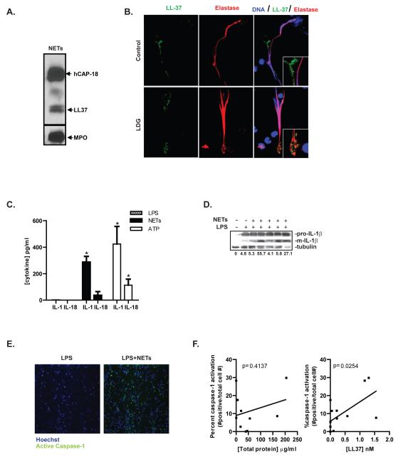
Neutrophil extracellular traps (NETs) represent an important defense mechanism against microorganisms. Clearance of NETs is impaired in a subset of patients with systemic lupus erythematosus, and NETosis is increased in neutrophils and, particularly, in low-density granulocytes derived from lupus patients. NETs are toxic to the endothelium, expose immunostimulatory molecules, activate plasmacytoid dendritic cells, and may participate in organ damage through incompletely characterized pathways. To better understand the role of NETs in fostering dysregulated inflammation, we examined inflammasome activation in response to NETs or to LL-37, an antibacterial protein externalized on NETs. Both NETs and LL-37 activate caspase-1, the central enzyme of the inflammasome, in both human and murine macrophages, resulting in release of active IL-1β and IL-18. LL-37 activation of the NLRP3 inflammasome utilizes P2X7 receptor-mediated potassium efflux. NET and LL-37-mediated activation of the inflammasome is enhanced in macrophages derived from lupus patients. In turn, IL-18 is able to stimulate NETosis in human neutrophils. These results suggest that enhanced formation of NETs in lupus patients can lead to increased inflammasome activation in adjacent macrophages. This leads to release of inflammatory cytokines that further stimulate NETosis, resulting in a feed-forward inflammatory loop that could potentially lead to disease flares and/or organ damage.


Figures

Figure 1
Figure 1. NETs activate caspase-1 and IL-1β and IL-18 release
A. Purified NETs were examined by Western analysis to confirm the presence of known NET-associated proteins. Myeloperoxidase, the LL-37 precursor h-CAP-18, and cleaved LL-37 are detected. B. NET formation was assessed in LPS-treated-control neutrophils and in unstimulated LDGs. Expression of LL-37 (green) in the NETs was assessed. Neutrophil elastase is shown in red. Results are representative of three separate experiments. Total magnification is 630×. C. Healthy control MØ (n=4) were primed with 100 ng/ml LPS for 4 h, followed by media change and a 2 h exposure to NETs derived from LPS-treated control neutrophils or from unstimulated lupus LDGs. Exposure to 5mM ATP for 2 h was used as a positive control. IL-1β and IL-18 were quantified by ELISA. Results are mean pg/mL + SEM of seven separate experiments completed in duplicate. D. Western analysis of combined whole-cell lysate and extracellular media following culture with LPS or LPS+NETs as in Figure 1C. Exposure to NET fractions results in increased mature (active) IL-1β. Percent of mature (m)IL-1β versus the house-keeping protein tubulin are shown below the graph. E. Human LPS-primed-control MØ were exposed to NETs as in figure 1C, for 1 h. The caspase-1-specific carboxyfluorescein tagged YVAD-FMK (green) was added to the culture to detect activated caspase-1. Cells were counterstained with Hoechst (blue) and imaged by fluorescent microscopy. Images are representative of 4 independent experiments, each performed in triplicate. Total magnification is 100×. F. Analysis of caspase-1 activation, determined as in Figure 1E, compared to total protein concentration (left) or concentration of LL-37 (right) present in the NETs. LDGs=Low Density Granulocytes. *=p<0.05.
Figure 2
Figure 2. LL-37 activates IL-1β and IL-18 release, preferentially in M1 versus M2 human and murine MØ, and this is not enhanced by complexing with DNA
A. Human MØ differentiated in the presence of GM-CSF (M1) or X-Vivo 15 (M2) were primed with LPS for 4 h, then treated with 20μM LL-37, 20μM inactive scrambled peptide (Scr) or 5 mM ATP for 2 h. Quantification of IL-1β and IL-18 in supernatants was performed by ELISA. B. Murine MØ differentiated in the presence of GM-CSF (M1) or M-CSF (M2) were primed with LPS for 4 h, then treated with 20μM mCRAMP or 5 mM ATP for 2 h. IL-1β was quantified in harvested supernatants by ELISA. C. Human M1 MØ were treated for 1 h with the caspase-1-specific carboxyfluorescein tagged YVAD-FMK (green) added after 1 h of various stimuli. Cells were counterstained with Hoechst and imaged by fluorescent microscopy. Percentage of caspase-1 activation (right) was determined by dividing the number of caspase-1 and Hoechst double-positive cells by the total number of Hoechst positive cells D. Human M1 MØ were treated as in 2A. Whole cell lysates (WCL) or extracellular media (ECM) were harvested and active caspase-1 p20 fragment was detected by Western Blot. Densitometric values of p20/p45 are shown below the image. E. Human M1 MØ were treated as in 2A, followed by 2 hour exposure to 20 μM LL-37 or to LL-37 precomplexed with DNA. IL-1β was quantified in supernatants by ELISA. Results represent mean + SEM, n=4-8 for each experiment; samples were run in duplicate. *=p<0.05, **=p<0.01, ***=p<0.001, ns=non-significant. Rx=treatment.
Figure 3
Figure 3. The NLRP3 inflammasome is required for mCRAMP activation of caspase-1 and for release of IL-1β
A. Murine BMDM, isolated from WT or from haplotype-matched mice deficient in NLRP3, ASC or caspase-1 were differentiated in the presence of GM-CSF for 7 days, followed by priming with 100 ng/ml LPS for 4 h. MØ were treated with 5mM ATP or 20μM mCRAMP (CMP) for 2 h and supernatants harvested for quantification by ELISA. B. Supernatants and cell lysates isolated from MØ treated as in A were analyzed for active caspase-1 (p20) content using Western analysis. Results represent mean + SEM for four separate experiments. *=p<0.05, **=p<0.01, ***=p<0.001. Rx=treatment.
Figure 4
Figure 4. LL-37 activation of the inflammasome requires P2×7R-mediated potassium efflux
A. Murine BMDM isolated from WT or haplotype-matched mice deficient in the P2×7R were differentiated in the presence of GM-CSF for 7 days, followed by priming with 100 ng/ml LPS for 4 h. MØ were then treated with 20μM mCRAMP (CMP) or 5mM ATP for 2 h before harvesting supernatants. B. Similarly-treated MØ were used to detect active caspase-1 by Western Blot. C. Human MØ (n=4) were differentiated for 7 days in the presence of GM-CSF, followed by stimulation with 5mM ATP or 20μM LL-37 in serum-free media with or without 100 mM KCl for 2 h. Release of IL-1β, IL-6 and IL-18 was assessed by ELISA. Results represent mean + SEM. *=p<0.05. ***=p<0.001. Rx=treatment.
Figure 5
Figure 5. Activation of caspase-1 by LL-37 and NETs is enhanced in SLE MØ
A. Equal number of monocytes isolated from healthy controls (n=7) or from individuals with SLE (n=8) were differentiated for 7 days in GM-CSF, primed with LPS for 4 h, followed by stimulation with various concentrations of LL-37 for 2 h. Cytokine concentrations of the supernatants were measured by ELISA. B. MØ from healthy controls (n=5) or from individuals with SLE (n=5) were similarly differentiated, followed by LPS-priming for 4 h. MØ were then treated for 2 h with NETs isolated from LPS-stimulated control neutrophils (n=4) or from unstimulated LDGs (n=5). Caspase-1 activation was detected via incubation with FLICA-YVAD-fmk during the last hour after NETs treatment. Cells were stained with Hoechst and imaged via fluorescent microscopy (right-representative images). Percentage of caspase-1 activation was determined as in figure 2C after normalization to baseline activation of LPS alone (left). Results from LDG and control NETs were pooled for analysis. C. Released IL-1β and IL-18 from macrophages treated as in B was determined by ELISA. *=p<0.05. **=p<0.01.
Figure 6
Figure 6. IL-18 induces NET formation
A. Neutrophils isolated from healthy controls were plated in the presence of the cell-impermeable dye Sytox Green, followed by exposure to PMA, LPS or graded concentrations of recombinant active human IL-18. NET release was detected via fluorescence emission. B. Neutrophils isolated from healthy controls were seeded onto polylysine-coated coverslips followed by exposure to LPS or graded concentrations of recombinant human IL-18. NETs were detected via staining for DNA (blue) and elastase (red). NETs were determined by co-localization of DNA and elastase and compared to the total number of neutrophils present to determine the percent of NETting neutrophils. *=p<0.05. C. Neutrophils isolated from healthy controls or LDGs isolated from lupus patients were plated in the presence of Sytox Green with or without a blocking antibody to the IL-18R (2μg/ml), IL-18 (3μg/ml) or an isotype-matched anti-IL-17R antibody (1μg/ml). Cells were then treated with IL-18 and NET release was detected via fluorescence emission. D. Healthy control macrophages were primed with LPS and treated with 25 ng/ml IL-18 or NETs isolated from IL-18-treated neutrophils (n=3). Percent of caspase-1 activation was detected and calculated as in figure 5B. E. Proposed pathway suggesting that NETs containing LL-37 can activate caspase-1 via the P2×7R. The subsequent processing and release of IL-1β and IL-18 further stimulate NETosis, leading to a self-perpetuating cycle of NET production and inflammasome activation. *=p<0.05. **=p<0.01, ***=p<0.001.

Similar articles

Cited by

References

    1. Brinkmann V, Zychlinsky A. Beneficial suicide: why neutrophils die to make NETs. Nat Rev Microbiol. 2007;5:577–582. - PubMed
    1. Kessenbrock K, Krumbholz M, Schonermarck U, Back W, Gross WL, Werb Z, Grone H-J, Brinkmann V, Jenne DE. Netting neutrophils in autoimmune small-vessel vasculitis. Nat Med. 2009;15:623–625. - PMC - PubMed
    1. Knight JS, Kaplan MJ. Lupus neutrophils: ‘NET’ gain in understanding lupus pathogenesis. Current opinion in rheumatology. 2012;24:441–450. - PubMed
    1. Lande R, Ganguly D, Facchinetti V, Frasca L, Conrad C, Gregorio J, Meller S, Chamilos G, Sebasigari R, Riccieri V, Bassett R, Amuro H, Fukuhara S, Ito T, Liu YJ, Gilliet M. Neutrophils Activate Plasmacytoid Dendritic Cells by Releasing Self-DNA-Peptide Complexes in Systemic Lupus Erythematosus. Sci Transl Med. 2011;3:73ra19. - PMC - PubMed
    1. Hakkim A, Furnrohr BG, Amann K, Laube B, Abed UA, Brinkmann V, Herrmann M, Voll RE, Zychlinsky A. Impairment of neutrophil extracellular trap degradation is associated with lupus nephritis. Proc Natl Acad Sci U S A. 2010;107:9813–9818. - PMC - PubMed

Publication types

MeSH terms