. 2013 Jan 8;110(2):648-53.
doi: 10.1073/pnas.1218667110. Epub 2012 Dec 24.

Activated protein C ameliorates diabetic nephropathy by epigenetically inhibiting the redox enzyme p66Shc

Affiliations

Activated protein C ameliorates diabetic nephropathy by epigenetically inhibiting the redox enzyme p66Shc

Fabian Bock et al. Proc Natl Acad Sci U S A. .

Abstract

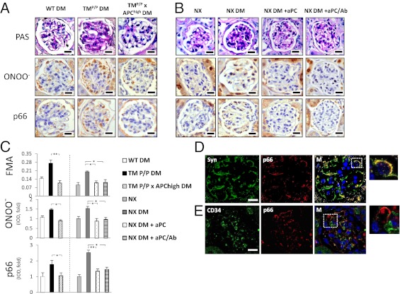
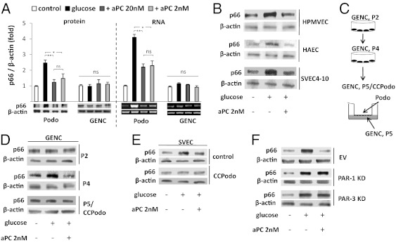
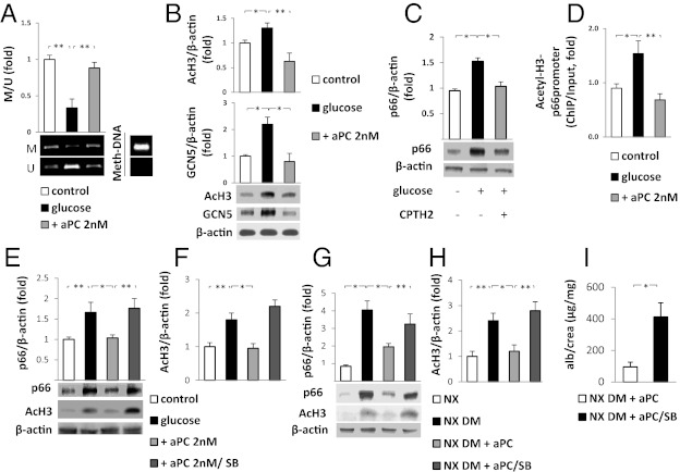
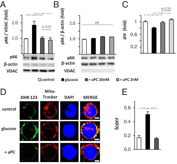
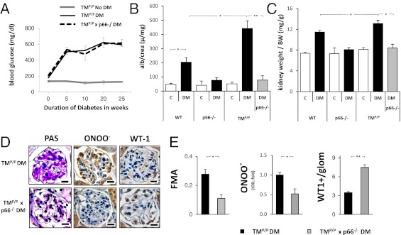
The coagulation protease activated protein C (aPC) confers cytoprotective effects in various in vitro and in vivo disease models, including diabetic nephropathy. The nephroprotective effect may be related to antioxidant effects of aPC. However, the mechanism through which aPC may convey these antioxidant effects and the functional relevance of these properties remain unknown. Here, we show that endogenous and exogenous aPC prevents glomerular accumulation of oxidative stress markers and of the redox-regulating protein p66(Shc) in experimental diabetic nephropathy. These effects were predominately observed in podocytes. In vitro, aPC inhibited glucose-induced expression of p66(Shc) mRNA and protein in podocytes (via PAR-1 and PAR-3) and various endothelial cell lines, but not in glomerular endothelial cells. Treatment with aPC reversed glucose-induced hypomethylation and hyperacetylation of the p66(Shc) promoter in podocytes. The hyperacetylating agent sodium butyrate abolished the suppressive effect of aPC on p66(Shc) expression both in vitro and in vivo. Moreover, sodium butyrate abolished the beneficial effects of aPC in experimental diabetic nephropathy. Inhibition of p66(Shc) expression and mitochondrial translocation by aPC normalized mitochondrial ROS production and the mitochondrial membrane potential in glucose-treated podocytes. Genetic ablation of p66(Shc) compensated for the loss of protein C activation in vivo, normalizing markers of diabetic nephropathy and oxidative stress. These studies identify a unique mechanism underlying the cytoprotective effect of aPC. Activated PC epigenetically controls expression of the redox-regulating protein p66(Shc), thus linking the extracellular protease aPC to mitochondrial function in diabetic nephropathy.


Conflict of interest statement

The authors declare no conflict of interest.

Figures

Fig. 1.
Fig. 1.
aPC prevents glucose-induced nitrotyrosine formation and p66Shc expression in podocytes. (A–C) Genetically compensating for aPC deficiency (TMP/P x APChigh DM mice) is sufficient to reverse enhanced extracellular matrix deposition (FMA), glomerular nitrotyrosine (ONOO) formation, and glomerular p66Shc expression in diabetic TMP/P (TMP/P DM) mice (A and C). In diabetic uninephrectomized wild-type mice (NX DM), therapeutic application of both native aPC (NX DM+aPC) or aPC lacking its anticoagulant properties (NX DM+aPC/Ab) reduced FMA, glomerular nitrotyrosine formation, and glomerular p66Shc expression to the levels observed in nondiabetic uninephrectomized wild-type mice (NX; B and C). Representative images of PAS, nitrotyrosine, and p66shc stained glomeruli (A and B) and bar graphs reflecting the overall results [C; integrated optical density (IOD) shown as the mean value ± SEM] are shown. *P < 0.05, **P < 0.005. p66Shc and nitrotyrosine were detected with HRP-DAB (brown) and hematoxylin counterstain (blue). (D and E) Representative confocal immunofluorescence images showing predominant colocalization (yellow) of p66Shc (red) with the podocyte marker synaptopodin (green; D), but not with the endothelial cell marker CD34 (green; E). Hoechst 33258 was used for nuclear counter staining (blue). (Scale bars: 20 µm.)
Fig. 2.
Fig. 2.
aPC prevents glucose-dependent p66Shc induction in podocytes in vitro. (A) Glucose (25 mM, 24 h) induces p66Shc protein and mRNA expression in mouse podocytes (Podo), but not in mouse glomerular endothelial cells (GENC). aPC prevents the glucose-mediated p66Shc induction in podocytes. Representative images of immunoblots (IB) and RT-PCR with bar graphs (>three independent repeat experiments; mean value ± SEM. *P < 0.05, **P < 0.005; ns, not significant). (B) Glucose-induced p66Shc expression in human pulmonary microvascular endothelial cells (HPMVEC), human aortic endothelial cells (HAEC), and the murine endothelial cell line SVEC4-10 is prevented by aPC. (C and D) Glucose induces p66Shc in late (P4) but not early (P2) passage GENCs. Coculture of P5 GENCs with murine podocytes (24 h, P5/CCPodo) renders GENCs once again unresponsive to glucose-induced expression of p66Shc. Scheme illustrating the experimental approach (C) and representative immunoblots (D). (E) Coculture of SVEC4-10 cells with mouse podocytes (24 h) prevents glucose-dependent p66Shc induction. (F) aPC-mediated inhibition of glucose-induced p66Shc expression requires PAR-1 and PAR-3. Representative immunoblots of p66Shc in control cells transfected with empty vector (EV) and in PAR-1 and PAR-3 knockdown (KD) podocytes.
Fig. 3.
Fig. 3.
aPC prevents glucose-induced p66Shc hypomethylation and H3 acetylation. (A) Glucose (25 mM) reduces methylation of the p66Shc promoter in podocytes, which is reversed by addition of 2 nM aPC. Representative image of methylated (M) and unmethylated (U) p66Shc promoter DNA revealed by methylation-specific -PCR(MSP) and a bar graph showing the ratio of methylated to unmethylated p66Shc promoter DNA (M/U, fold change) are shown. Universal methylated mouse DNA (Meth-DNA) was used as a control. (B) Glucose (25 mM)-induced H3 acetylation (AcH3) and increased H3 acetyltransferase (GCN5) expression is prevented by the addition of aPC in podocytes. (C) Treatment of podocytes with the GCN5 inhibitor CPTH2 (50 µM) is sufficient to prevent glucose-induced p66Shc expression. (D) H3 acetylation within the p66Shc promoter is induced by glucose and prevented by aPC (2 nM). Acetylated H3 was immunoprecipitated with an AcH3 antibody, and the p66shc promoter was quantitated by qRT-PCR (ChIP data were normalized to input). (E and F) Sodium butyrate (SB) increases H3 acetylation even in the presence of aPC (+aPC 2nM/SB) and abolishes the suppressive effects of aPC on H3 acetylation and p66Shc expression in glucose-treated podocytes (25 mM glucose). (G and H) The suppressive effect of aPC on H3 acetylation and p66Shc expression in the renal cortex of uninephrectomized diabetic wild-type mice (NX DM+aPC) is abolished by concomitant treatment with SB (NX DM+aPC/SB). (I) SB abolishes the aPC-mediated reduction in albuminuria in uninephrectomized wild-type diabetic mice (NX DM+aPC/SB). Representative images of MSP (A) or immunoblots (B, C, E, and G) and bar graphs (mean value ± SEM) summarizing the results of at least three repeat experiments or five different mice are shown. *P < 0.05; **P < 0.005.
Fig. 4.
Fig. 4.
aPC prevents glucose-induced mitochondrial translocation of p66Shc, maintains mitochondrial membrane potential, and reduces mitochondrial ROS-generation in podocytes. (A and B) Glucose-induced (25 mM, 24 h) translocation of p66Shc into mitochondria is efficiently prevented by aPC. Representative immunoblots of p66Shc, VDAC (mitochondrial marker), and β-actin (cytosolic marker) in mitochondrial (A) or cytosolic (B) cellular subfractions. (C) Glucose reduces the MMP (Mito-Probe JC-1) in podocytes. aPC treatment of glucose-exposed podocytes maintains the MMP. (D and E) Representative fluorescence microscopy images showing single mouse podocytes (D). Podocytes were left untreated (5 mM glucose) or stimulated with glucose (25 mM, 24 h) without or with aPC (20 nM). ROS formation was monitored by using dihydrorhodamine (DHR, green) and localized to mitochondria by using Mitotracker CMX (red). Hoechst 33258 was used for nuclear counter staining (blue). Bar graph (E) summarizing the results (mean value ± SEM) of four independent repeat experiments using automated digital colocalization analyses, yielding the Icorr index. p66, p66Shc; *P < 0.05 and **P < 0.005; ns, not significant. (Scale bars: 5 µm.)
Fig. 5.
Fig. 5.
Genetic p66Shc deficiency compensates for the loss of TM-dependent PC activation in experimental diabetic nephropathy. (A) No difference in the blood glucose levels was apparent between diabetic TMP/P and diabetic TMP/P x p66Shc−/− mice. (B and C) p66Shc deficiency normalizes albuminuria (B) and kidney weight (C) in mice with genetically impaired PC activation (TMP/P mice). (D and E) p66Shc deficiency in diabetic TMP/P mice reduces histological indices of diabetic nephropathy. PAS staining, fractional mesangial area (FMA; E), and immunohistochemical analyses of nitrotyrosine and the podocyte protein WT-1 are shown. Representative images (D) and bar graphs summarizing the IODs (E). Data are presented as the mean ± SEM; at least six mice per group (AC) or ≥30 glomeruli per genotype and mouse (D and E) were analyzed. *P < 0.05, **P < 0.005. (Scale bars: 20 µm.)

Similar articles

Cited by

References

    1. Griffin JH, Zlokovic BV, Mosnier LO. Protein C anticoagulant and cytoprotective pathways. Int J Hematol. 2012;95(4):333–345. - PMC - PubMed
    1. Riewald M, Petrovan RJ, Donner A, Mueller BM, Ruf W. Activation of endothelial cell protease activated receptor 1 by the protein C pathway. Science. 2002;296(5574):1880–1882. - PubMed
    1. Joyce DE, Gelbert L, Ciaccia A, DeHoff B, Grinnell BW. Gene expression profile of antithrombotic protein c defines new mechanisms modulating inflammation and apoptosis. J Biol Chem. 2001;276(14):11199–11203. - PubMed
    1. Zhong Z, et al. Activated protein C therapy slows ALS-like disease in mice by transcriptionally inhibiting SOD1 in motor neurons and microglia cells. J Clin Invest. 2009;119(11):3437–3449. - PMC - PubMed
    1. Yamaji K, et al. Activated protein C, a natural anticoagulant protein, has antioxidant properties and inhibits lipid peroxidation and advanced glycation end products formation. Thromb Res. 2005;115(4):319–325. - PubMed

Publication types

MeSH terms