

Desmoplastic melanoma: a review
- PMID: 23267722
- PMCID: PMC4703041
- DOI: 10.1016/j.jaad.2012.10.041
Desmoplastic melanoma: a review
Abstract
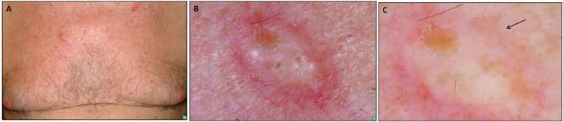
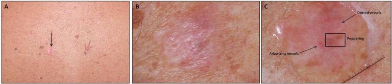
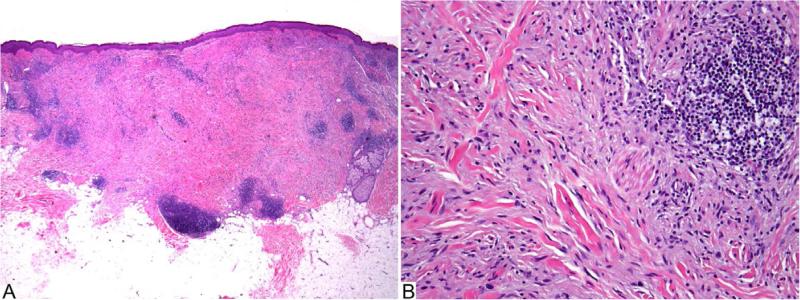
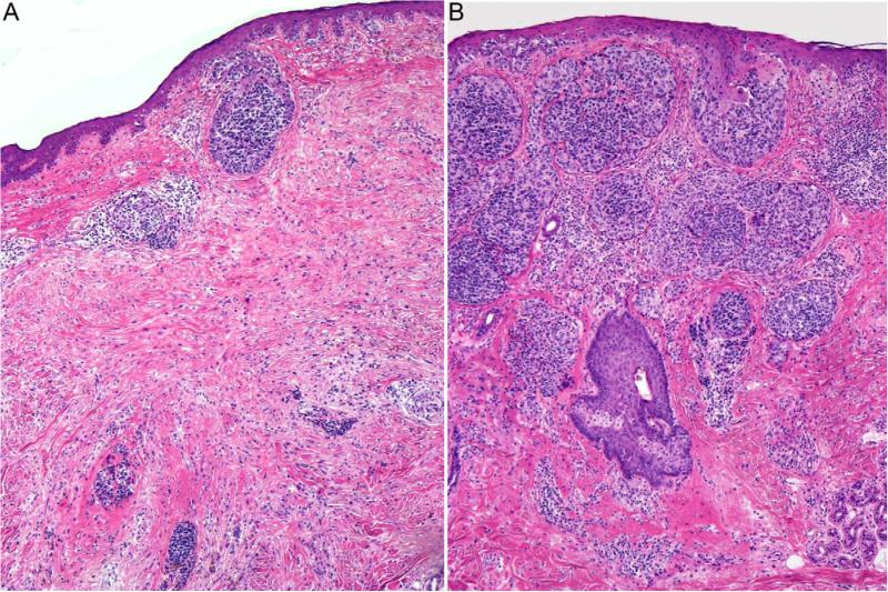
Desmoplastic melanoma (DM) is a variant of spindle cell melanoma typically found on chronically sun-damaged skin of older individuals. Early diagnosis can be challenging because it is often amelanotic and has a predominantly dermal component. DM can be difficult to diagnose not only clinically but also histologically, and can be mistaken for a variety of benign and malignant nonmelanocytic spindle cell tumors when viewed on prepared histopathology slides. Pathologists have observed that DMs can manifest significant variation with respect to the extent of intratumoral cellularity, fibrosis, and/or perineural invasion. Furthermore, some tumors present with a pure desmoplastic invasive component (>90%) while other tumors display mixed features of DM and nondesmoplastic melanoma. This has led to the separation of DM into 2 histologic subtypes, pure and mixed. With a focus on the distinction between pure and mixed DM, this review will detail what is currently known about the diagnostic features of DM, discuss risk and prognostic factors, and examine the current literature on disease progression and management.
Copyright © 2013 American Academy of Dermatology, Inc. Published by Mosby, Inc. All rights reserved.
Figures
References
-
- Quinn MJ, Crotty KA, Thompson JF, Coates AS, O'Brien CJ, McCarthy WH. Desmoplastic and desmoplastic neurotropic melanoma: experience with 280 patients. Cancer. 1998;83:1128–35. - PubMed
-
- Feng Z, Wu X, Chen V, Velie E, Zhang Z. Incidence and survival of desmoplastic melanoma in the United States, 1992-2007. J Cutan Pathol. 2011;38:616–24. - PubMed
-
- Conley J, Lattes R, Orr W. Desmoplastic malignant melanoma (a rare variant of spindle cell melanoma). Cancer. 1971;28:914–36. - PubMed
-
- Labrecque PG, Hu CH, Winkelmann RK. On the nature of desmoplastic melanoma. Cancer. 1976;38:1205–13. - PubMed
-
- Bernaba BN, Vogiatzis PI, Binder SW, Cassarino DS. Potentially useful markers for desmoplastic melanoma: an analysis of KBA.62, p-AKT, and ezrin. Am J Dermatopathol. 2011;33:333–7. quiz 8-40. - PubMed
Publication types
MeSH terms
Grants and funding
LinkOut - more resources
Full Text Sources
Other Literature Sources
Medical
