. 2013 Mar:14:406-25.
doi: 10.1016/j.meegid.2012.12.002. Epub 2012 Dec 23.

A survey of host range genes in poxvirus genomes

Affiliations

A survey of host range genes in poxvirus genomes

Kirsten A Bratke et al. Infect Genet Evol. 2013 Mar.

Abstract

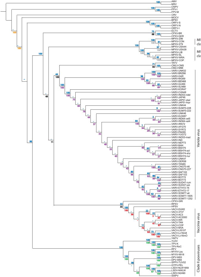
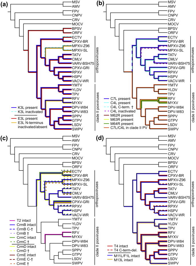
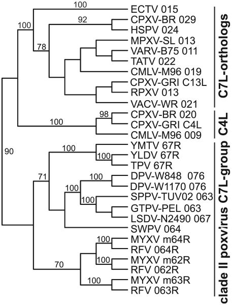
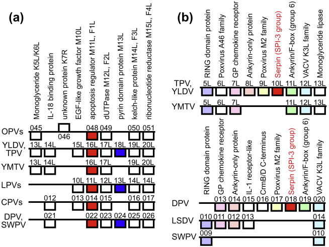
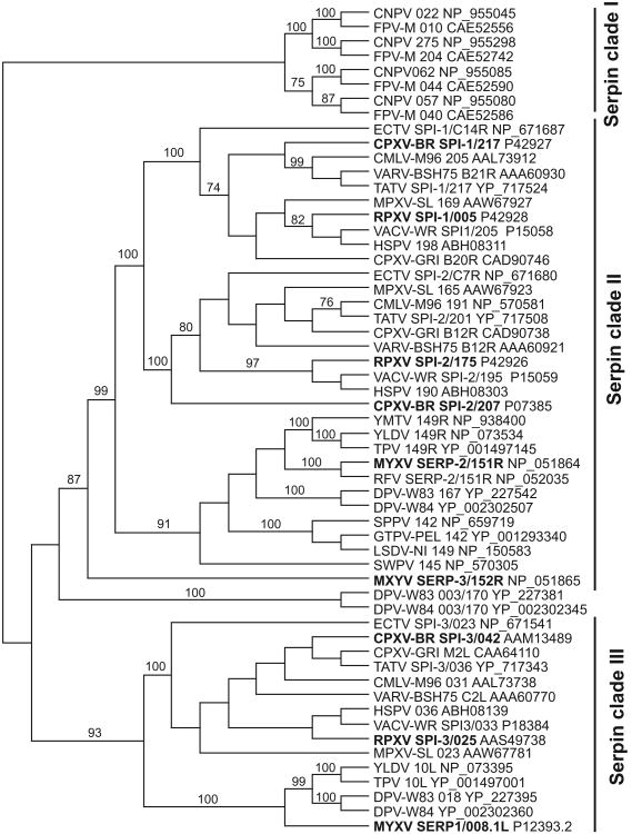
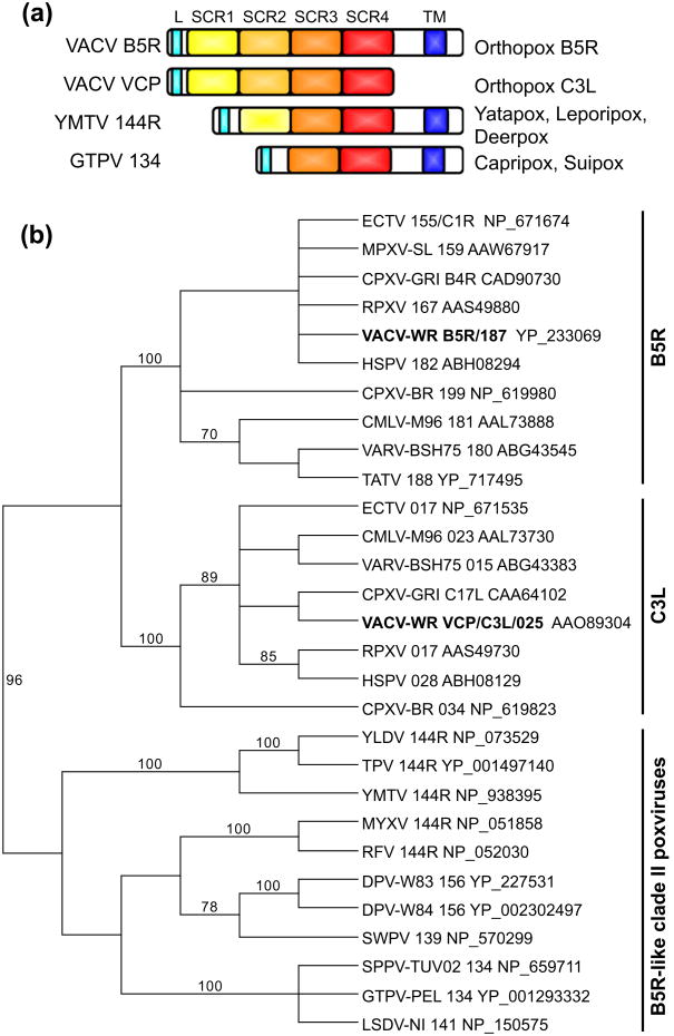
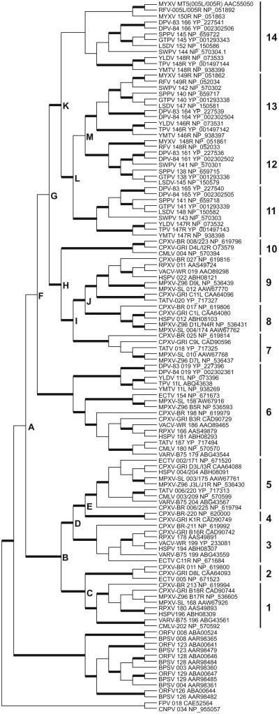
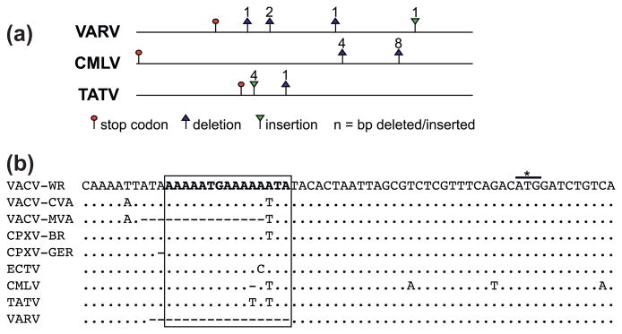
Poxviruses are widespread pathogens, which display extremely different host ranges. Whereas some poxviruses, including variola virus, display narrow host ranges, others such as cowpox viruses naturally infect a wide range of mammals. The molecular basis for differences in host range are poorly understood but apparently depend on the successful manipulation of the host antiviral response. Some poxvirus genes have been shown to confer host tropism in experimental settings and are thus called host range factors. Identified host range genes include vaccinia virus K1L, K3L, E3L, B5R, C7L and SPI-1, cowpox virus CP77/CHOhr, ectromelia virus p28 and 022, and myxoma virus T2, T4, T5, 11L, 13L, 062R and 063R. These genes encode for ankyrin repeat-containing proteins, tumor necrosis factor receptor II homologs, apoptosis inhibitor T4-related proteins, Bcl-2-related proteins, pyrin domain-containing proteins, cellular serine protease inhibitors (serpins), short complement-like repeats containing proteins, KilA-N/RING domain-containing proteins, as well as inhibitors of the double-stranded RNA-activated protein kinase PKR. We conducted a systematic survey for the presence of known host range genes and closely related family members in poxvirus genomes, classified them into subgroups based on their phylogenetic relationship and correlated their presence with the poxvirus phylogeny. Common themes in the evolution of poxvirus host range genes are lineage-specific duplications and multiple independent inactivation events. Our analyses yield new insights into the evolution of poxvirus host range genes. Implications of our findings for poxvirus host range and virulence are discussed.


Figures

Fig. 1
Fig. 1
Phylogenetic tree of completely sequenced poxviruses. A neighbor-joining tree based on 28 core protein families forms the backbone for this tree (blue bootstraps). Concatenated alignments of larger numbers of protein families present in single copy in subsets of the 104 genomes are used to resolve poorly supported branches. Bootstrap values in different colors correspond to these sub-trees. Orange: 36 families in CRV, MOCV, BPSV, FPV-I, AMV. Green: 95 families in clade II. Black: 83 families in orthopoxviruses. Grey: 128 families in MPXV. Purple: 114 families in VARV. Red: 100 families in VACV, HSPV, RPXV. Schematic tree topology is shown and branch lengths are not drawn to scale. (For interpretation of the references to color in this figure legend, the reader is referred to the web version of this article.)
Fig. 2
Fig. 2
Presence of selected poxvirus host range genes projected on trees showing phylogenetic relationships of representative poxvirus strains. (a) Presence of VACV K3L orthologs is indicated above the branches (dark blue for deduced functional genes, dashed light blue lines represent inactivated genes). VACV E3L orthologs are shown below the branches in red (full length genes) and yellow (N-terminus inactivated). (b) Presence of genes of the C7L/M63R family is indicated. Solid blue lines above the branches represent C7L orthologs in orthopoxviruses, C4L orthologs are shown below branches as solid light blue lines (intact ORFs), dashed light blue lines (C-terminus inactivated) or dashed purple lines (ORFs inactivated). C7L/C4L orthologs in clade II poxviruses are shown above the branches, except for leporipoxviruses, for which three homologs (M62R, M63R and M64R) are present. (c) Presence of Tumor necrosis factor receptor II homologs is shown. CrmB orthologs are shown in solid blue lines (intact genes), dashed blue lines (C-terminus inactivated) or dashed light purple lines (ORF inactivated). CrmC orthologs are shown as solid (intact genes) or dashed green lines (ORFs inactivated). CrmD orthologs are represented by solid (intact genes) or dashed yellow lines (ORFs inactivated). CrmE orthologs are shown in solid red lines (intact genes), dashed red lines (C-terminus inactivated) or dashed organge lines (ORF inactivated). Purple lines indicate presence of T4. (d) Occurrence of T4, M11L/F1L and M13L families is shown. Presence of T4 genes is indicated by solid (intact genes) and dashed (C-terminus deleted) red lines. Blue and yellow lines indicate presence of M11L/F1L and M13L orthologs, respectively. Schematic tree topologies are shown and branch lengths are not drawn to scale. (For interpretation of the references to color in this figure legend, the reader is referred to the web version of this article.)
Fig. 3
Fig. 3
Phylogenetic relationship of C7L/M063R family genes. The phylogenetic relationship between C7L/M063R homologs is shown. A mid-point rooted tree was generated from a multiple protein sequence alignment of the indicated sequences using the neighbor-joining method. Bootstrap support of ≥70 is indicated above the branches.
Fig. 4
Fig. 4
Synteny conservation around F1L/M11L, M13L and SPI-3 genes in poxvirus genomes. (a) Synteny conservation around F1L/M11L and M13L in selected poxvirus genomes is shown. Boxes above and below horizontal lines represent genes, transcribed from left to right or right to left, respectively. Homologous genes in different genomes are aligned vertically, with any functional information and the name of the gene (in vaccinia virus Copenhagen and/or myxoma virus as a leporipoxvirus (LPV) representative listed at the top. F1L/M11L orthologs are highlighted in red; M13L orthologs are shown in blue. Gene names are given for representative family members: for orthopoxviruses (OPVs), gene names for CPXV-BR are shown. Note that CPXV-BR ORF 045 encoding a lipase is disrupted in VACV (K5L/K6L) and CMLV by multiple in/dels, and 95% of it is deleted in VARV. For capripoxviruses (CPVs), gene names for GTPV and SSPV are shown. The same genes are also present in LSDV but are represented by ORFs 015-020. Identical gene content for this region is found in DPV and SWPV. ORFs (021-026) found in DPV are shown here, and correspond to ORFs 011-016 in SWPV. (b) Synteny conservation around SPI-3 orthologs in selected clade II poxvirus genomes is shown. SPI-3 orthologs are highlighted in red. Identical colors in the upper and lower part of this figure represent orthologous genes in respective genomes. The gene that corresponds to LSDV ORF13 is disrupted in SPPV and GTPV by multiple indels. (For interpretation of the references to color in this figure legend, the reader is referred to the web version of this article.)
Fig. 5
Fig. 5
Phylogenetic relationship of poxvirus serpin proteins. The midpoint-rooted phylogenetic tree was constructed using the neighbor-joining method with a multiple protein sequence alignment of the indicated sequences. Bootstrap support of ≥70 is indicated above the branches. Prototypic poxvirus serpins are highlighted (bold letters). Names of proteins/ORFs and accession numbers are indicated.
Fig. 6
Fig. 6
Structure and phylogeny of the B5R/VCP family. (a) Schematic representation of the domain organization of B5R and VCP (C3L) family proteins. Leader sequences (light blue), transmembrane domains (dark blue) and short complement-like repeats (SCR1-4: yellow, light orange, orange and red, respectively) are highlighted. (b) Mid-point rooted neighbor-joining tree of a protein multiple sequence alignment of SCR3 and SCR4 from indicated sequences. Bootstrap support of ≥70 is indicated above the branches. Names of proteins/ORFs and accession numbers are indicated. (For interpretation of the references to color in this figure legend, the reader is referred to the web version of this article.)
Fig. 7
Fig. 7
Phylogeny of poxvirus ANK/F-box proteins. Neighbor-joining analyses performed with multiple protein sequence alignments generated using Muscle, from which non-alignable portions were excised using GBlocks. The tree was rooted to FPV 018 and CNPV 034. Bold lines mark branches with bootstrap values of ≥80 in all analyses performed. Letters A–M indicate inferred duplication events. Orthopoxvirus and clade II PV ANK/F-box proteins were classified into 14 different groups depending on phylogenetic clades.
Fig. 8
Fig. 8
Gene-inactivating mutations in K1L orthologs of variola, camelpox and taterapox viruses. (a) Horizontal lines represent genes of VARV, CMLV and TATV that are orthologous to vaccinia virus K1L. Inactivating mutations are indicated by red circles (stop codons), deletions (blue triangles) and insertions (inverse green triangles). Numbers indicate deleted or inserted nucleotides, which lead to frame shifts. (b) Comparison of the 5′ region of K1L orthologs in different orthopoxviruses. The box highlights an early promoter core motif that is found at position −42 to −28, relative to the predicted start codon (asterisk). Dots indicate nucleotides that are identical to VACV-WR. Dashes indicate deletions. (For interpretation of the references to color in this figure legend, the reader is referred to the web version of this article.)

Similar articles

Cited by

References

    1. Afonso CL, Tulman ER, Delhon G, Lu Z, Viljoen GJ, Wallace DB, Kutish GF, Rock DL. Genome of crocodilepox virus. J Virol. 2006;80:4978–4991. - PMC - PubMed
    1. Afonso CL, Tulman ER, Lu Z, Zsak L, Kutish GF, Rock DL. The genome of fowlpox virus. J Virol. 2000;74:3815–3831. - PMC - PubMed
    1. Alejo A, Ruiz-Arguello MB, Ho Y, Smith VP, Saraiva M, Alcami A. A chemokine-binding domain in the tumor necrosis factor receptor from variola (smallpox) virus. Proc Natl Acad Sci USA. 2006;103:5995–6000. - PMC - PubMed
    1. Ali AN, Turner PC, Brooks MA, Moyer RW. The SPI-1 gene of rabbitpox virus determines host range and is required for hemorrhagic pock formation. Virology. 1994;202:305–314. - PubMed
    1. Altschul SF, Madden TL, Schaffer AA, Zhang J, Zhang Z, Miller W, Lipman DJ. Gapped BLAST and PSI-BLAST: a new generation of protein database search programs. Nucleic Acids Res. 1997;25:3389–3402. - PMC - PubMed

Publication types