Review
. 2013:6:47-62.
doi: 10.1109/RBME.2012.2233468. Epub 2012 Dec 20.

The expanding world of tissue engineering: the building blocks and new applications of tissue engineered constructs

Affiliations
Review

The expanding world of tissue engineering: the building blocks and new applications of tissue engineered constructs

Pinar Zorlutuna et al. IEEE Rev Biomed Eng. 2013.

Abstract

The field of tissue engineering has been growing in the recent years as more products have made it to the market and as new uses for the engineered tissues have emerged, motivating many researchers to engage in this multidisciplinary field of research. Engineered tissues are now not only considered as end products for regenerative medicine, but also have emerged as enabling technologies for other fields of research ranging from drug discovery to biorobotics. This widespread use necessitates a variety of methodologies for production of tissue engineered constructs. In this review, these methods together with their non-clinical applications will be described. First, we will focus on novel materials used in tissue engineering scaffolds; such as recombinant proteins and synthetic, self assembling polypeptides. The recent advances in the modular tissue engineering area will be discussed. Then scaffold-free production methods, based on either cell sheets or cell aggregates will be described. Cell sources used in tissue engineering and new methods that provide improved control over cell behavior such as pathway engineering and biomimetic microenvironments for directing cell differentiation will be discussed. Finally, we will summarize the emerging uses of engineered constructs such as model tissues for drug discovery, cancer research and biorobotics applications.


Figures

Figure 1
Figure 1
Control of cell spreading by reversibly crosslinkable ECM components, by selectively crosslinking, uncrosslinking and re-crosslinking the alginate component within the collagen hydrogel, cell shape can be controlled in 3D. Cells were labeled for actin fibers with AlexiFluor488-Phalloidin (Adapted from [22]).
Figure 2
Figure 2
Hexamer polypeptides with a hydrophilic head and a hydrophobic tail can self- assemble into millimeter scale fibers. A) Schematic model of the hexamer polypeptide sequences (LIVAGD and AIVAGD) that assemble into fibers via α-helical pairing. B) Fiber mats and single fibers obtained from the self-assembling polypeptides, these fibers can support several cell types such as mesenchymal stem cells and epithelial cells (Adapted from [44]).
Figure 3
Figure 3
A) Schematics of the modular construct design and fabrication. HepG2 cell encapsulated cylindrical collagen gels were seeded with human umbilical-vein endothelial cells (HUVECs). After HUVECs completely covered the gel surface, which takes usually 2 to 3 days, the HUVEC-seeded cylinders can be assembled into a larger structure to form the modular construct that can be used to perfuse supply nutrients to the cells through formed network of interconnected channels. B) Light microscopy images of collagen–HepG2 module prior to HUVEC seeding. C) On Day 7 of the culture period, HUVECs on the surface of the modules was stained for VE-cadherin and imaged using confocal microscopy to show the confluent layer of HUVECs on the surface of the modules. D) A flow circuit was used to perfuse PBS or media the modular construct. E) After media perfusion of 7 days, collagen–HepG2–HUVEC modules were retrieved and imaged using confocal microscopy (Adapted from [58]).
Figure 4
Figure 4
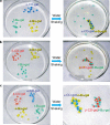
Macroscale assembly of gel modules through selective molecular recognition of acrylamide gels modified with cyclodextrin or hydrocarbon groups. A) Selective assembly of α-CD-gel (blue) and n-Bu-gel (yellow) upon shaking in water in the presence of t-Bu-gel (dark green). B) Selective assembly of β-CD-gel (red) and t-Bu-gel (dark green) upon shaking in water in the presence of n-Bu-gel (yellow). C) Selective assembly of α-CD-gel/n-Bu-gel and β-CD-gel/t-Bu-gel upon shaking in water in the presence of all gel types (Adapted from [70]).
Figure 5
Figure 5
Fabrication of temperature responsive substrates for generating cell aggregates. A-B) PNIPAAm microwells were fabricated by soft lithography, then cells were seeded and retrieved after aggregate formation. Phase contrast and fluorescent microscopy images of the aggregates released from C) PNIPAAm compared to D) PEG microwells (Adapted from [89]).
Figure 6
Figure 6
A) Schematics of the simultaneous protein immobilization using 2 photon laser scanning method. A femtosecond laser was used to immobilize maleimide-barnase, represented with the black circle, followed by a wash step to remove unbound maleimide-barnase. Then, maleimide-streptavidin was immobilized, represented by orange square, and again followed by a wash step. After this step, a protein of interest which has been fused to barstar and biotin was introduced. They will specifically bind to barnase and streptavidin, respectively. B and C) Confocal microscopy images of simultaneous patterning of biotin–CNTF (red) and barstar–SHH (green) (Scale bar: 100 μm) (Adapted from [153]).
Figure 7
Figure 7
Non-linear microscopy techniques SHG and CARS allow simultaneous, real-time monitoring of cell-fibrous scaffold (bacterial cellulose) interactions. A) Interactions of vascular smooth muscle cells with nanofibrillar bacterial cellulose was monitored at specific time points to quantify cell proliferation, migration and ECM secretion B) Secreted collagen fibers can be distinguished from the cellulose fibers due to the substantial difference in their average fiber size (A collagen fiber is indicated with an arrow around vascular smooth muscle cells) (Adapted from [206]).

References

    1. Engler AJ, et al. Matrix Elasticity Directs Stem Cell Lineage Specification. Cell. 2006 Aug;126:677–689. - PubMed
    1. O'Brien FJ, et al. The effect of pore size on cell adhesion in collagen-GAG scaffolds. Biomaterials. 2005 Feb;26:433–441. - PubMed
    1. Khademhosseini A, et al. Progress in Tissue Engineering. Scientific American. 2009 May;300:64–71. - PubMed
    1. Lazic T, Falanga V. Bioengineered Skin Constructs and Their Use in Wound Healing. Plastic and Reconstructive Surgery. 2011 Jan;127:75S–90S. - PubMed
    1. Roberts SJ, et al. Clinical applications of musculoskeletal tissue engineering. British Medical Bulletin. 2008 Jun;86:7–22. - PubMed

Publication types