

doi: 10.1097/WOX.0b013e3181e2eb2e.
Epub 2010 Jun 15.
Dermatology for the allergist
Affiliations
- PMID: 23268431
- PMCID: PMC3488899
- DOI: 10.1097/WOX.0b013e3181e2eb2e
Item in Clipboard
Dermatology for the allergist
World Allergy Organ J.
2010 Jun.
Abstract
Allergists/immunologists see patients with a variety of skin disorders. Some, such as atopic and allergic contact dermatitis, are caused by abnormal immunologic reactions, whereas others, such as seborrheic dermatitis or rosacea, lack an immunologic basis. This review summarizes a select group of dermatologic problems commonly encountered by an allergist/immunologist.
Conflict of interest statement
The authors state that they have no conflicts of interest to declare.
Figures
Atopic dermatitis. Note erythema and scaling of flexural areas. Reprinted with permission from Habif, Clinical Dermatology, 5th ed. Elsevier, 2009.
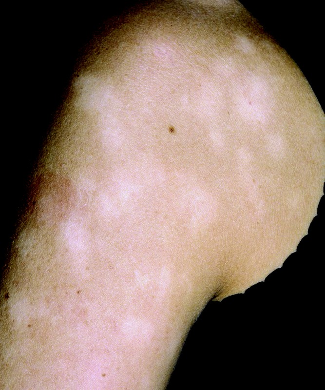
Pityriasis alba hypopigmentation. Reprinted with permission from Habif, Clinical Dermatology, 5th ed. Elsevier, 2009.
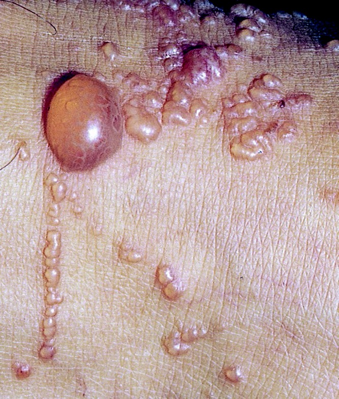
Poison ivy rash with vesicles, blisters, and linear lesions. Reprinted with permission from Habif, Clinical Dermatology, 5th ed. Elsevier, 2009.
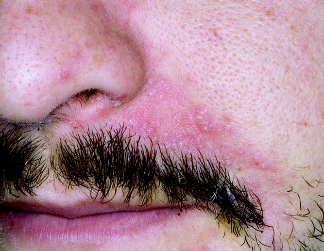
Seborrhoic dermatitis affecting nasolabial folds. Reprinted with permission from Habif, Clinical Dermatology, 5th ed. Elsevier, 2009.
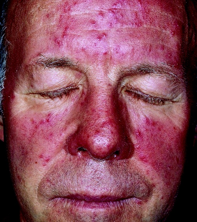
Rosacea erythema and few pustules affecting the face. Reprinted with permission from Habif, Clinical Dermatology, 5th ed. Elsevier, 2009.
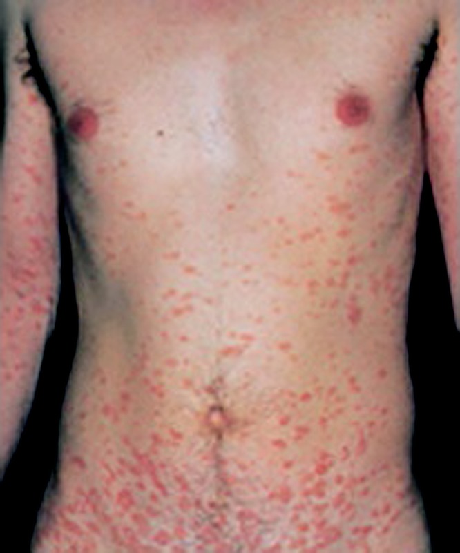
Pityriasis rosea fully evolved two weeks after onset. Reprinted with permission from Habif, Clinical Dermatology, 4th ed. Elsevier, 2004.
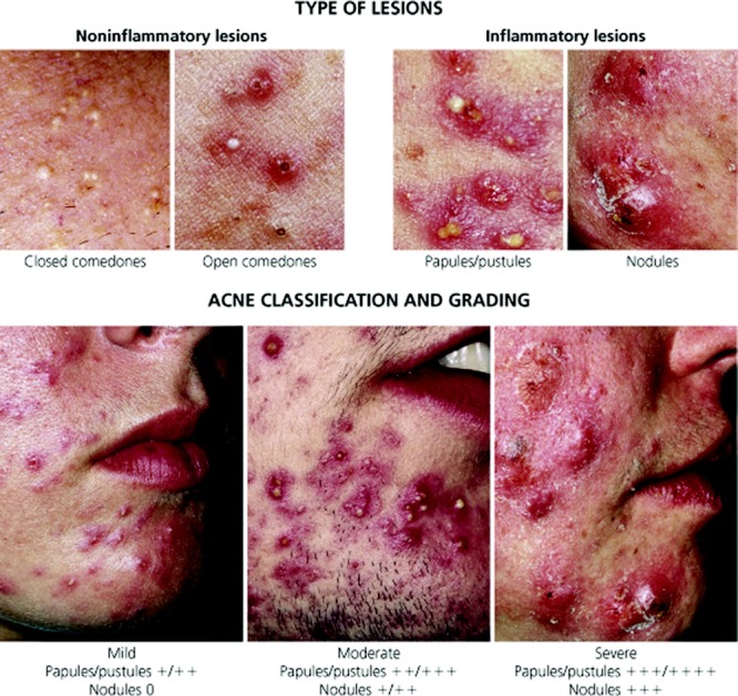
Acne classification and grading. Habif, Clinical Dermatology, 5th ed. Elsevier, 2009.
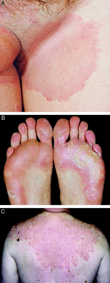
A, Tinea cruris; B, tinea pedis; C, tinea corporis. Reprinted with permission from Habif, Clinical Dermatology. 4th ed. Elsevier, 2004; 5th ed, Elsevier, 2009.
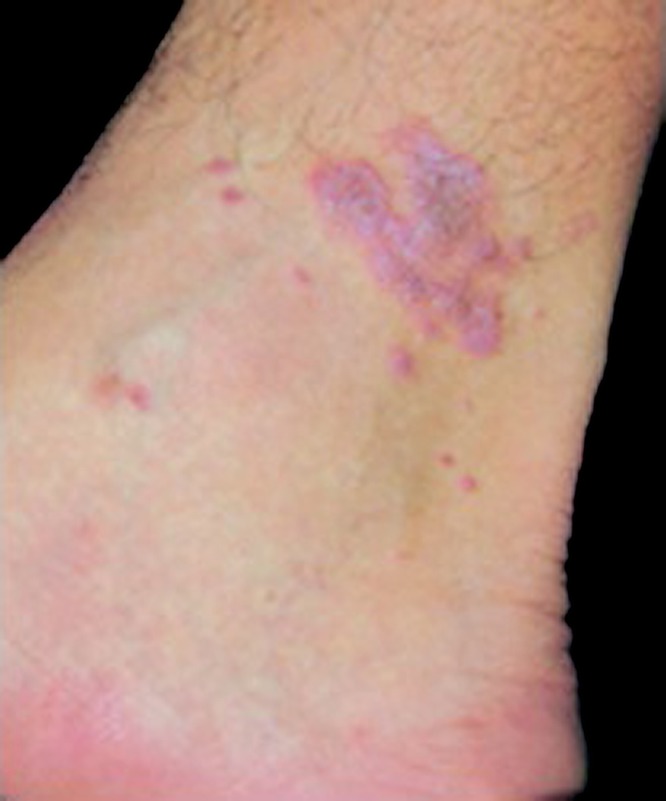
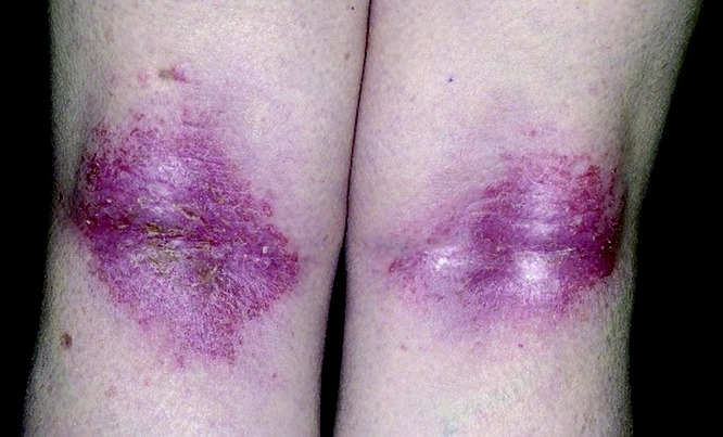
Lichen planus with characteristic purple papules and Wickham's striae. Habif, Clinical Dermatology, 4th ed. Elsevier, 2004.
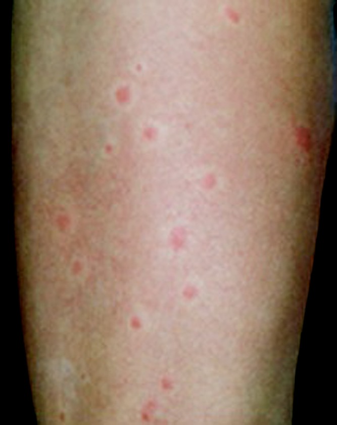
Ictus reaction from insect bites. Habif, Clinical Dermatology, 4th ed. Elsevier, 2004.
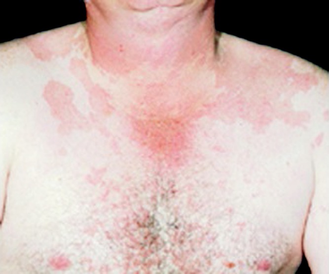
Tinea versicolor of neck and trunk appears as confluent, fawn-colored patches. Habif, Clinical Dermatology, 4th ed. Elsevier, 2004.
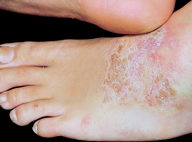
Lichen simplex chronicus (neurodermatitis) caused by chronic scratching by the opposite heel. Habif, Clinical Dermatology, 4th ed. Elsevier, 2004.
Similar articles
-
Dermatologic Problems Commonly Seen by the Allergist/Immunologist.J Allergy Clin Immunol Pract. 2020 Jan;8(1):102-112. doi: 10.1016/j.jaip.2019.07.019. Epub 2019 Jul 25. J Allergy Clin Immunol Pract. 2020. PMID: 31351991 Review.
-
Electronic health records-Applications for the allergist/immunologist: All that glitters is not gold.Allergy Asthma Proc. 2016 Jul;37(4):273-8. doi: 10.2500/aap.2016.37.3959. Allergy Asthma Proc. 2016. PMID: 27401314 Review.
-
Long-term management of moderate-to-severe adult atopic dermatitis: a consensus by the Italian Society of Dermatology and Venereology (SIDeMaST), the Association of Italian Territorial and Hospital Allergists and Immunologists (AAIITO), the Italian Association of Hospital Dermatologists (ADOI), the Italian Society of Allergological, Environmental and Occupational Dermatology (SIDAPA), and the Italian Society of Allergy, Asthma and Clinical Immunology (SIAAIC).Ital J Dermatol Venerol. 2022 Feb;157(1):1-12. doi: 10.23736/S2784-8671.21.07129-2. Epub 2021 Dec 21. Ital J Dermatol Venerol. 2022. PMID: 34929995
-
Photocontact Dermatitis and Its Clinical Mimics: an Overview for the Allergist.Clin Rev Allergy Immunol. 2019 Feb;56(1):32-40. doi: 10.1007/s12016-018-8696-x. Clin Rev Allergy Immunol. 2019. PMID: 29951786 Review.
-
Contact allergens for the allergist.Ann Allergy Asthma Immunol. 2022 Jun;128(6):629-644. doi: 10.1016/j.anai.2022.03.022. Epub 2022 Mar 25. Ann Allergy Asthma Immunol. 2022. PMID: 35346877 Review.
Cited by
-
Attenuation Effect of Radiofrequency Irradiation on UV-B-Induced Skin Pigmentation by Decreasing Melanin Synthesis and through Upregulation of Heat Shock Protein 70.Molecules. 2021 Dec 17;26(24):7648. doi: 10.3390/molecules26247648. Molecules. 2021. PMID: 34946730 Free PMC article.
-
Radiofrequency Irradiation Attenuates High-Mobility Group Box 1 and Toll-like Receptor Activation in Ultraviolet B-Induced Skin Inflammation.Molecules. 2021 Feb 28;26(5):1297. doi: 10.3390/molecules26051297. Molecules. 2021. PMID: 33670841 Free PMC article.
-
Not All Fun and Games: A Case Report of Contact Dermatitis Related to Slime and Play-Doh.Cureus. 2020 Sep 6;12(9):e10272. doi: 10.7759/cureus.10272. Cureus. 2020. PMID: 33042709 Free PMC article.
-
Antifungal Resistance in Dermatology.Indian J Dermatol. 2018 Sep-Oct;63(5):361-368. doi: 10.4103/ijd.IJD_131_17. Indian J Dermatol. 2018. PMID: 30210155 Free PMC article. Review.
-
Characterization of IgE-binding proteins in the salivary glands of Simulium nigrogilvum (Diptera: Simuliidae).Parasitol Res. 2019 Aug;118(8):2353-2359. doi: 10.1007/s00436-019-06383-x. Epub 2019 Jul 1. Parasitol Res. 2019. PMID: 31263951
References
-
- Laughter D, Istvan JA, Tofte SJ, Hanifin JM. The prevalence of atopic dermatitis in Oregon schoolchildren. J Am Acad Dermatol. 2000;43:649–655 - PubMed
-
- Georg R. Atopic Dermatitis. London: Saunders; 1975
-
- Uehara M, Kimura C. Descendant family history of atopic dermatitis. Acta Derm Venereol. 1993;73:62–63 - PubMed
-
- McGrath JA, Uitto J. The filaggrin story: novel insights into skin-barrier function and disease. Trends Mol Med. 2008;14:20–27 - PubMed
-
- Vasilopoulos Y, Cork MJ, Murphy R, Williams HC, Robinson DA, et al. Genetic association between an AACC insertion in the 3′UTR of the stratum corneum chymotryptic enzyme gene and atopic dermatitis. J Invest Dermatol. 2004;123:62–66 - PubMed
LinkOut - more resources
Full Text Sources
Other Literature Sources
