. 2013 Feb 1;41(3):1807-16.
doi: 10.1093/nar/gks1282. Epub 2012 Dec 24.

A novel mechanism of eukaryotic translation initiation that is neither m7G-cap-, nor IRES-dependent

Affiliations

A novel mechanism of eukaryotic translation initiation that is neither m7G-cap-, nor IRES-dependent

Ilya M Terenin et al. Nucleic Acids Res. .

Abstract

Resistance of translation of some eukaryotic messenger RNAs (mRNAs) to inactivation of the cap-binding factor eIF4E under unfavorable conditions is well documented. To date, it is the mechanism of internal ribosome entry that is predominantly thought to underlay this stress tolerance. However, many cellular mRNAs that had been considered to contain internal ribosome entry sites (IRESs) failed to pass stringent control tests for internal initiation, thus raising the question of how they are translated under stress conditions. Here, we show that inserting an eIF4G-binding element from a virus IRES into 5'-UTRs of strongly cap-dependent mRNAs dramatically reduces their requirement for the 5'-terminal m(7)G-cap, though such cap-independent translation remains dependent on a vacant 5'-terminus of these mRNAs. Importantly, direct binding of eIF4G to the 5'-UTR of mRNA makes its translation resistant to eIF4F inactivation both in vitro and in vivo. These data may substantiate a new paradigm of translational control under stress to complement IRES-driven mechanism of translation.


Figures

Figure 1.
Figure 1.
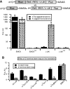
Direct interaction of eIF4G with 5′-UTRs of mRNAs dramatically affects cap dependence of their translation. (A) Schematic representation of mRNAs used in the experiments. The black partition represents vector-derived nucleotides. (B) Comparison of translation efficiencies of the indicated m7G- and A-capped monocistronic mRNAs in Krebs-2 cytoplasmic extract. The translation level of m7G-capped mRNA with the β-globin 5′-UTR is set to 1. (C) Results of transfection of the same mRNAs into HEK293T cells. Fluc/Rluc value for m7G-capped mRNA with the β-globin 5′-UTR is arbitrarily set to 1. (D) Translation in HEK293T cells lysate. The translation level of m7G-capped mRNA with the β-globin 5′-UTR is set to 1. (E) Transfection in BHK21 cells. Fluc/Rluc value for m7G-capped mRNA with the β-globin 5′-UTR is arbitrarily set to 1. (F) Indicated A- or m7G-capped mRNAs lacking poly(A)-tail were translated in Krebs-2 cells S30 extract. The translation level of A-capped mRNA with the L1JK 5′-UTR is set to 1. (G) Translation in a WGE. The translation level of A-capped mRNA with the L1JK 5′-UTR is set to 1. All translation and transfection experiments were repeated at least in triplicate.
Figure 2.
Figure 2.
Despite binding of eIF4G at internal positions of mRNAs, the translation is 5′ end-dependent. (A) m7G-capped bicistronic or equivalent equimolar amounts of m7G-capped Rluc with A-capped Fluc mRNAs were transfected into HEK293T cells. Plotted are relative values of the Firefly luciferase expression. The Fluc value for the bicistronic EMCVmut mRNA is arbitrarily set to 1. (B) Results of translation of indicated mRNAs with or without a 5′-terminal stem-loop in Krebs-2 cytoplasmic extract. The Fluc/Rluc value for m7G-capped mRNA with the β-globin 5′-UTR and the stem-loop is arbitrarily set to 1. Error bars represent standard error of the mean.
Figure 3.
Figure 3.
Translation driven by the CITE is initiated by ribosomal scanning. (A) Real-time analysis of firefly luciferase accumulation in Krebs-2 extract for A-capped mRNAs with different 5′-UTR lengths (indicated on the plot). (B) Full translation times for the m7G- and A-capped mRNAs JK-Fluc, ΔL1JK-Fluc and L1JK-Fluc as calculated from the kinetic curves.
Figure 4.
Figure 4.
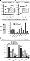
Effect of eIF4E inactivation on the translation of various mRNA constructs in vitro in Krebs-2 extract and HEK293T cells. (A) An example of the effect of exogenous 4E-BP1 on the translation of the m7G- and A-capped monocistronic L1JK-Fluc mRNAs containing either an active or disabled binding site for eIF4G. (B) Effect of the addition of m7GTP on the translation of the same mRNAs. (C) Effect of eIF4G cleavage by poliovirus 2A protease on the translation of mRNAs with various 5′-UTRs. The translation level of m7G-capped β-globin mRNA is set to 1. The left panel shows western blot analysis of eIF4G without or with cleavage by 2A protease. (D) HEK293T cells were depleted of serum (24 h before transfection), treated with wortmannin (2 h before transfection) or treated with etoposide (36 h before transfection). Then, cells were transfected with the indicated mRNAs, and the expression of reporters was analyzed 3 h later. The data are presented as a ratio of expression from the mRNA with the intact JK-domain to that of the mRNA with the mutated J-K domain for the L1JK-Fluc mRNA or JK-βGlo-Fluc mRNA. The ratio of Fluc/Rluc values of mRNAs with or without intact J-K domain in untreated cells is set to 1. Error bars represent standard error of the mean.
Figure 5.
Figure 5.
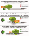
Model of translation mediated by enhancers of cap-independent translation. There are two ways how a 43 S pre-initiation complex can enter the 5′-terminus of an mRNA. The first way is mediated by interaction of the m7G-cap with eIF4E and can be inhibited in various ways. The second one is mediated by an internal element that binds eIF4G. The captured 43 S pre-initiation complex irrespective of the presence of the m7G-cap is then either actively transferred to the 5′-terminus of the mRNA or being snapped in the internal position catches the 5′-end, where it can initiate scanning. In the third mechanism, IRES forces a ribosome to enter mRNA directly into the internal positions of the latter.

References

    1. Sonenberg N, Hinnebusch AG. Regulation of translation initiation in eukaryotes: mechanisms and biological targets. Cell. 2009;136:731–745. - PMC - PubMed
    1. Gingras AC, Raught B, Sonenberg N. eIF4 initiation factors: effectors of mRNA recruitment to ribosomes and regulators of translation. Annu. Rev. Biochem. 1999;68:913–963. - PubMed
    1. Andreev DE, Dmitriev SE, Terenin IM, Prassolov VS, Merrick WC, Shatsky IN. Differential contribution of the m7G-cap to the 5' end-dependent translation initiation of mammalian mRNAs. Nucleic Acids Res. 2009;37:6135–6147. - PMC - PubMed
    1. Gilbert WV. Alternative ways to think about cellular internal ribosome entry. J. Biol. Chem. 2010;285:29033–29038. - PMC - PubMed
    1. Kozak M. A second look at cellular mRNA sequences said to function as internal ribosome entry sites. Nucleic Acids Res. 2005;33:6593–6602. - PMC - PubMed

Publication types

MeSH terms