

Review
doi: 10.1056/NEJMra1208129.
Mechanisms and management of retinopathy of prematurity
Affiliations
- PMID: 23268666
- PMCID: PMC3695731
- DOI: 10.1056/NEJMra1208129
Item in Clipboard
Review
Mechanisms and management of retinopathy of prematurity
N Engl J Med.
.
No abstract available
Figures
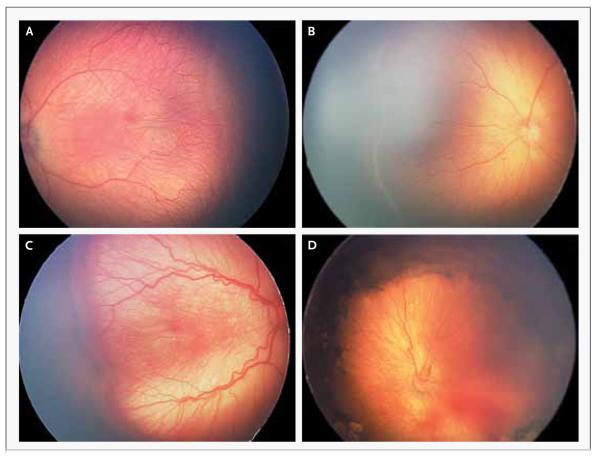
The images were obtained with a neonatal retinal imaging system (RetCam, Clarity Medical Systems). Panel A shows a line between the vascularized and avascularized retina (stage 1). Panel 2 shows a ridge between the vascularized and avascularized retina (stage 2). Panel 3 shows a thickened ridge with aberrant intravitreal angiogenesis (stage 3). Panel 4 shows partial retinal detachment (stage 4), which is seen best at the right side of the image where the underlying choroidal vascular detail is out of focus.
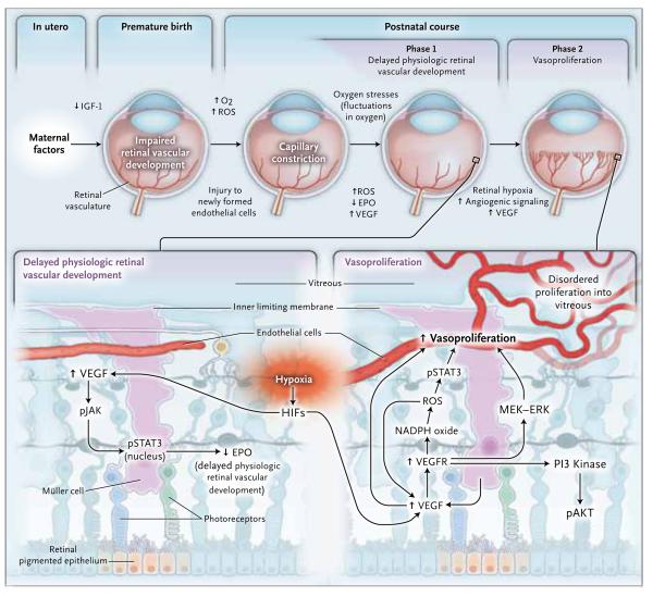
In retinopathy of prematurity in the United States today, there is initially delayed physiologic retinal vascular development, resulting in a peripheral avascular area of the retina (phase 1). Later, vasoproliferation in the form of intravitreal angiogenesis can occur at the junction of avascularized and vascularized retina (phase 2). EPO denotes erythropoietin, ERK extracellular signal-regulated kinase, HIF hypoxia-inducible factor, IGF-1 insulin-like growth factor 1, MEK mitogen-activated protein–ERK, O2 oxygen, pAKT phosphorylated protein kinase B, PI3 phosphatidylinositol 3, pJAK phosphorylated Janus kinase, pSTAT3 phosphorylated signal transducer and activator of transcription 3, ROS reactive oxygen species, VEGF vascular endothelial growth factor, and VEGFR vascular endothelial growth factor receptor.
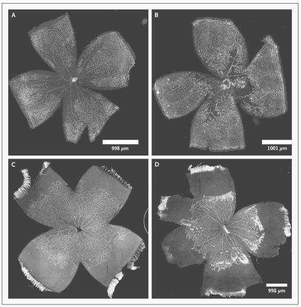
The center of the cloverleaf is the optic nerve, and the farthest extent of each leaf of the cloverleaf is the ora serrata. There is no macula in the mouse or rat retina. The panels on the left show phase 1 retinopathy of prematurity, and the panels on the right show phase 2 retinopathy of prematurity. In Panel A, a p12 mouse model shows central hyperoxia-induced vaso-obliteration after 5 days of 75% oxygen. In Panel B, a p17 mouse model after an additional 5 days in room air shows vasoproliferation at the junctions of the vascularized and avascularized retina. This model may represent retinopathy of prematurity in the United States in the 1950s or possibly in regions lacking resources to provide oxygen regulation and monitoring. In Panel C, a p14 rat model shows delayed physiologic retinal vascular development with peripheral avascularized retina after seven cycles of fluctuations between 50% and 10% oxygen. In Panel D, a p18 rat model after 4 days in ambient air shows vasoproliferation at the junction of the vascularized and avascularized retina; this model represents retinopathy of prematurity as currently seen in the United States and other countries where oxygen is regulated.
Comment in
-
Mechanisms and management of retinopathy of prematurity.N Engl J Med. 2013 Mar 21;368(12):1162-3. doi: 10.1056/NEJMc1301021. N Engl J Med. 2013. PMID: 23514302 No abstract available.
-
Mechanisms and management of retinopathy of prematurity.N Engl J Med. 2013 Mar 21;368(12):1161. doi: 10.1056/NEJMc1301021. N Engl J Med. 2013. PMID: 23514303 No abstract available.
-
Mechanisms and management of retinopathy of prematurity.N Engl J Med. 2013 Mar 21;368(12):1161-2. doi: 10.1056/NEJMc1301021. N Engl J Med. 2013. PMID: 23514304 No abstract available.
References
-
- Good WV, Hardy RJ, Dobson V, et al. The incidence and course of retinopathy of prematurity: findings from the Early Treatment for Retinopathy of Prematurity Study. Pediatrics. 2005;116:15–23. - PubMed
-
- Lad EM, Hernandez-Boussard T, Morton JM, Moshfeghi DM. Incidence of retinopathy of prematurity in the United States: 1997 through 2005. Am J Ophthalmol. 2009;148:451–8. - PubMed
-
- Section on Ophthalmology American Academy of Pediatrics. American Academy of Ophthalmology. American Association for Pediatric Ophthalmology and Strabismus Screening examination of premature infants for retinopathy of prematurity. Pediatrics. 2006;117:572–6. - PubMed
- Pediatrics. 2006;118:1324. Erratum.
-
- Terry TL. Extreme prematurity and fibroblastic overgrowth of persistent vascular sheath behind each crystalline lens: Preliminary report. Am J Ophthalmol. 1942;25:203–4. - PubMed
-
- Michaelson IC. The mode of development of the vascular system of the retina with some observations on its significance for certain retinal diseases. Trans Ophthalmol Soc UK. 1948;68:137–80.
Publication types
MeSH terms
Substances
Grants and funding
LinkOut - more resources
Full Text Sources
Other Literature Sources