The neurovascular unit as a selective barrier to polymorphonuclear granulocyte (PMN) infiltration into the brain after ischemic injury
- PMID: 23269317
- PMCID: PMC3578720
- DOI: 10.1007/s00401-012-1076-3
The neurovascular unit as a selective barrier to polymorphonuclear granulocyte (PMN) infiltration into the brain after ischemic injury
Abstract
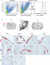
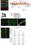
The migration of polymorphonuclear granulocytes (PMN) into the brain parenchyma and release of their abundant proteases are considered the main causes of neuronal cell death and reperfusion injury following ischemia. Yet, therapies targeting PMN egress have been largely ineffective. To address this discrepancy we investigated the temporo-spatial localization of PMNs early after transient ischemia in a murine transient middle cerebral artery occlusion (tMCAO) model and human stroke specimens. Using specific markers that distinguish PMN (Ly6G) from monocytes/macrophages (Ly6C) and that define the cellular and basement membrane boundaries of the neurovascular unit (NVU), histology and confocal microscopy revealed that virtually no PMNs entered the infarcted CNS parenchyma. Regardless of tMCAO duration, PMNs were mainly restricted to luminal surfaces or perivascular spaces of cerebral vessels. Vascular PMN accumulation showed no spatial correlation with increased vessel permeability, enhanced expression of endothelial cell adhesion molecules, platelet aggregation or release of neutrophil extracellular traps. Live cell imaging studies confirmed that oxygen and glucose deprivation followed by reoxygenation fail to induce PMN migration across a brain endothelial monolayer under flow conditions in vitro. The absence of PMN infiltration in infarcted brain tissues was corroborated in 25 human stroke specimens collected at early time points after infarction. Our observations identify the NVU rather than the brain parenchyma as the site of PMN action after CNS ischemia and suggest reappraisal of targets for therapies to reduce reperfusion injury after stroke.
Figures


Similar articles
-
The temporo-spatial localization of polymorphonuclear cells related to the neurovascular unit after transient focal cerebral ischemia.Brain Res. 2014 Oct 24;1586:184-92. doi: 10.1016/j.brainres.2014.08.037. Epub 2014 Aug 22. Brain Res. 2014. PMID: 25152468
-
Carcinoembryonic antigen-related cell adhesion molecule 1 inhibits MMP-9-mediated blood-brain-barrier breakdown in a mouse model for ischemic stroke.Circ Res. 2013 Sep 27;113(8):1013-22. doi: 10.1161/CIRCRESAHA.113.301207. Epub 2013 Jun 18. Circ Res. 2013. PMID: 23780386
-
Hydrogen peroxide pretreatment of perfused canine vessels induces ICAM-1 and CD18-dependent neutrophil adherence.Circulation. 1991 Nov;84(5):2154-66. doi: 10.1161/01.cir.84.5.2154. Circulation. 1991. PMID: 1682068
-
Role of polymorphonuclear neutrophils in the reperfused ischemic brain: insights from cell-type-specific immunodepletion and fluorescence microscopy studies.Ther Adv Neurol Disord. 2018 Sep 14;11:1756286418798607. doi: 10.1177/1756286418798607. eCollection 2018. Ther Adv Neurol Disord. 2018. PMID: 30245743 Free PMC article. Review.
-
Roles of Polymorphonuclear Neutrophils in Ischemic Brain Injury and Post-Ischemic Brain Remodeling.Front Immunol. 2022 Jan 11;12:825572. doi: 10.3389/fimmu.2021.825572. eCollection 2021. Front Immunol. 2022. PMID: 35087539 Free PMC article. Review.
Cited by
-
Central nervous system zoning: How brain barriers establish subdivisions for CNS immune privilege and immune surveillance.J Intern Med. 2022 Jul;292(1):47-67. doi: 10.1111/joim.13469. Epub 2022 Mar 14. J Intern Med. 2022. PMID: 35184353 Free PMC article.
-
Exploring Key Genes and Mechanisms in Respiratory Syncytial Virus-Infected BALB/c Mice via Multi-Organ Expression Profiles.Front Cell Infect Microbiol. 2022 May 2;12:858305. doi: 10.3389/fcimb.2022.858305. eCollection 2022. Front Cell Infect Microbiol. 2022. PMID: 35586251 Free PMC article.
-
Day 1 neutrophil-to-lymphocyte ratio (NLR) predicts stroke outcome after intravenous thrombolysis and mechanical thrombectomy.Front Neurol. 2022 Aug 9;13:941251. doi: 10.3389/fneur.2022.941251. eCollection 2022. Front Neurol. 2022. PMID: 36016545 Free PMC article.
-
Neutrophil extracellular traps released by neutrophils impair revascularization and vascular remodeling after stroke.Nat Commun. 2020 May 19;11(1):2488. doi: 10.1038/s41467-020-16191-y. Nat Commun. 2020. PMID: 32427863 Free PMC article.
-
Thromboinflammatory challenges in stroke pathophysiology.Semin Immunopathol. 2023 May;45(3):389-410. doi: 10.1007/s00281-023-00994-4. Epub 2023 Jun 5. Semin Immunopathol. 2023. PMID: 37273022 Free PMC article. Review.
References
-
- Agrawal S, Anderson P, Durbeej M, van Rooijen N, Ivars F, Opdenakker G, Sorokin LM. Dystroglycan is selectively cleaved at the parenchymal basement membrane at sites of leukocyte extravasation in experimental autoimmune encephalomyelitis. J Exp Med. 2006;203:1007–1019. doi: 10.1084/jem.20051342. - DOI - PMC - PubMed
-
- Arumugam TV, Salter JW, Chidlow JH, Ballantyne CM, Kevil CG, Granger DN. Contributions of LFA-1 and Mac-1 to brain injury and microvascular dysfunction induced by transient middle cerebral artery occlusion. Am J Physiol Heart Circ Physiol. 2004;287:H2555–H2560. doi: 10.1152/ajpheart.00588.2004. - DOI - PubMed

