Left ventricular and aortic dysfunction in cystic fibrosis mice
- PMID: 23269368
- PMCID: PMC4170835
- DOI: 10.1016/j.jcf.2012.11.012
Left ventricular and aortic dysfunction in cystic fibrosis mice
Abstract
Background: Left ventricular (LV) abnormalities have been reported in cystic fibrosis (CF); however, it remains unclear if loss of cystic fibrosis transmembrane conductance regulator (CFTR) function causes heart defects independent of lung disease.
Methods: Using gut-corrected F508del CFTR mutant mice (ΔF508), which do not develop human lung disease, we examined in vivo heart and aortic function via 2D transthoracic echocardiography and LV catheterization.
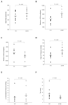
Results: ΔF508 mouse hearts showed LV concentric remodeling along with enhanced inotropy (increased +dP/dt, fractional shortening, decreased isovolumetric contraction time) and greater lusitropy (-dP/dt, Tau). Aortas displayed increased stiffness and altered diastolic flow. β-adrenergic stimulation revealed diminished cardiac reserve (attenuated +dP/dt,-dP/dt, LV pressure).
Conclusions: In a mouse model of CF, CFTR mutation leads to LV remodeling with alteration of cardiac and aortic functions in the absence of lung disease. As CF patients live longer, more active lives, their risk for cardiovascular disease should be considered.
Keywords: Aorta; CFTR; Cystic fibrosis; Left ventricular function.
Copyright © 2012 European Cystic Fibrosis Society. Published by Elsevier B.V. All rights reserved.
Figures


References
-
- Nagel G, Hwang TC, Nastiuk KL, Nairn AC, Gadsby DC. The protein kinase A-regulated cardiac Cl− channel resembles the cystic fibrosis transmembrane conductance regulator. Nature. 1992;360:81–4. - PubMed
-
- Opie LH. The Heart: Physiology, from Cell to Circulation. Lippencott-Raven; Philadelphia, PA: 1998.
-
- Duan DY, Liu LL, Bozeat N, Huang ZM, Xiang SY, Wang GL, et al. Functional role of anion channels in cardiac diseases. Acta Pharmacologica Sinica. 2005;26:265–78. - PubMed
-
- Kuzumoto M, Takeuchi A, Nakai H, Oka C, Noma A, Matsuoka S. Simulation analysis of intracellular Na+ and Cl− homeostasis during beta 1-adrenergic stimulation of cardiac myocyte. Prog Biophys Mol Biol. 2008;96:171–86. - PubMed
Publication types
MeSH terms
Substances
Grants and funding
LinkOut - more resources
Full Text Sources
Other Literature Sources
Medical
Molecular Biology Databases
Research Materials

