. 2013 Feb 8;288(6):3907-17.
doi: 10.1074/jbc.M112.413906. Epub 2012 Dec 26.

Site-specific N-linked glycosylation of receptor guanylyl cyclase C regulates ligand binding, ligand-mediated activation and interaction with vesicular integral membrane protein 36, VIP36

Affiliations

Site-specific N-linked glycosylation of receptor guanylyl cyclase C regulates ligand binding, ligand-mediated activation and interaction with vesicular integral membrane protein 36, VIP36

Najla Arshad et al. J Biol Chem. .

Abstract

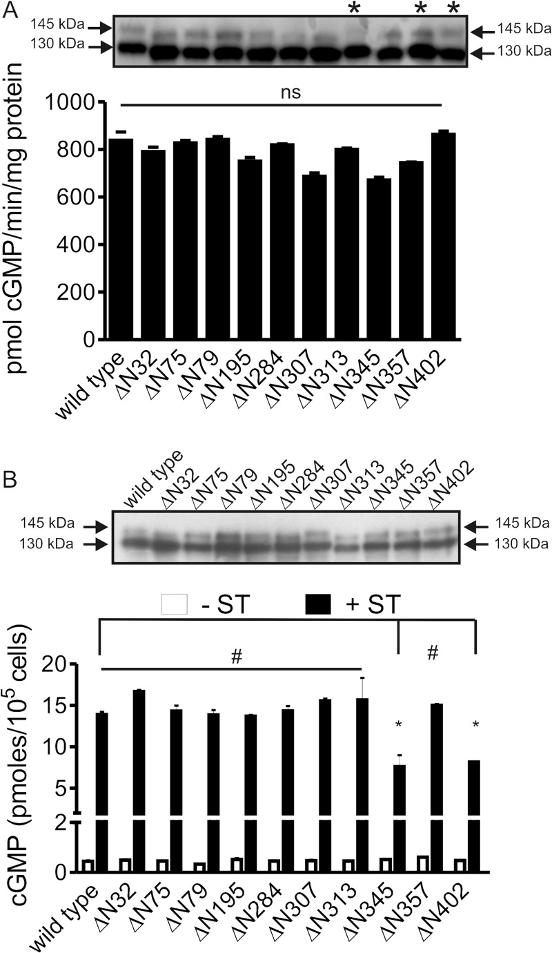
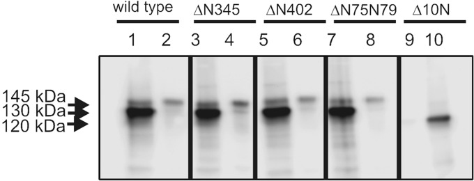
Guanylyl cyclase C (GC-C) is a multidomain, membrane-associated receptor guanylyl cyclase. GC-C is primarily expressed in the gastrointestinal tract, where it mediates fluid-ion homeostasis, intestinal inflammation, and cell proliferation in a cGMP-dependent manner, following activation by its ligands guanylin, uroguanylin, or the heat-stable enterotoxin peptide (ST). GC-C is also expressed in neurons, where it plays a role in satiation and attention deficiency/hyperactive behavior. GC-C is glycosylated in the extracellular domain, and differentially glycosylated forms that are resident in the endoplasmic reticulum (130 kDa) and the plasma membrane (145 kDa) bind the ST peptide with equal affinity. When glycosylation of human GC-C was prevented, either by pharmacological intervention or by mutation of all of the 10 predicted glycosylation sites, ST binding and surface localization was abolished. Systematic mutagenesis of each of the 10 sites of glycosylation in GC-C, either singly or in combination, identified two sites that were critical for ligand binding and two that regulated ST-mediated activation. We also show that GC-C is the first identified receptor client of the lectin chaperone vesicular integral membrane protein, VIP36. Interaction with VIP36 is dependent on glycosylation at the same sites that allow GC-C to fold and bind ligand. Because glycosylation of proteins is altered in many diseases and in a tissue-dependent manner, the activity and/or glycan-mediated interactions of GC-C may have a crucial role to play in its functions in different cell types.


Figures

FIGURE 1.
FIGURE 1.
Effect of glycosylation on GC-C activity. A, monolayers of HEK293T cells were transfected with wild type GC-C or GC-CΔ10N and 18 h post-transfection and treated with 20 μg/ml of tunicamycin as indicated for 24 h. Western blot analysis of membrane protein (40 μg) was performed using GC-C:C8 monoclonal antibody. B, receptor binding analysis was carried out using membrane protein (40 μg) as described in the text. Data shown are mean ± S.E. from a representative experiment, with experiments performed twice with duplicate determinations. C, in vitro guanylyl cyclase assays using MnGTP as substrate was performed with membrane protein (5 μg) from cells expressing the wild type or GC-CΔ10N receptors, untreated or treated with tunicamycin. Data shown are mean ± S.E. from a representative experiment, with experiments performed thrice with duplicate determinations. D, receptor binding assays and Scatchard analyses was performed using equivalent amounts of GST-tagged wild type or GC-CΔ10N ECDs bound to glutathione beads, purified from E. coli. Experiments were performed three times, and values represent the mean ± S.E. across the experiments. E, membrane protein (40 μg) from cells expressing GC-C were subjected to deglycosylation using PNGase F, followed by Western blot analysis with GC-C:C8 monoclonal antibody. F, in vitro ST-mediated cGMP production was measured in control or deglycosylated membrane protein (10 μg) with MgGTP as substrate. Data shown are mean ± S.E. from a representative experiment, with experiments performed thrice, with duplicate determinations. Statistical significance was evaluated using the Student's t test. *, p < 0.001; ns, not significant in comparison with the wild type receptor.
FIGURE 2.
FIGURE 2.
Analysis of site-specific mutants of the 10 predicted glycosylation sites in GC-C. A, membrane protein (5 μg) from cells expressing the indicated GC-C mutants were assayed for guanylyl cyclase activity in the presence of MnGTP as substrate. Data shown are mean ± S.E. from a representative experiment, with experiments performed twice with duplicate determinations. Inset, membrane protein (10 μg) from cells expressing the indicated mutants was analyzed by Western blotting using GC-C:C8 monoclonal antibody. *, mutants that did not show a mobility shift. Data are representative of experiments performed three times. No significant (ns) difference was seen in guanylyl cyclase activity across experiments, when activity was normalized to the level of expression of the mutant proteins. B, ST (10−7 m) was applied to cultures of cells expressing wild type or the indicated mutant forms of GC-C and cGMP produced measured by radioimmunoassay. Data shown are mean ± S.E. from a representative assay with duplicate determinations, and assays were repeated three times. Statistical significance was evaluated using the Student's t test. *, p < 0.01; #, p > 0.05 compared with wild type GC-C. Inset shows a Western blot of lysates prepared from transfected cells of a representative transfection.
FIGURE 3.
FIGURE 3.
Role of glycosylation at Asn402. A, Western blot analysis performed with wild type and indicated mutant receptors using the GC-C:C8 monoclonal antibody. A representative blot is shown of experiments repeated three times. B, ST (10−7 m) was applied to cultures of cells expressing wild type or mutant forms of GC-C and cGMP produced measured by radioimmunoassay. Data shown are mean ± S.E. from a representative assay with duplicate determinations, and assays were repeated three times. C, in vitro ST-mediated cGMP production was measured with membrane protein (10 μg) from cells expressing the indicated GC-C mutant using MgGTP as substrate. Data shown are mean ± S.E. from a representative experiment, with experiments performed twice, with duplicate determinations. Statistical significance was evaluated using the Student's t test. *, p < 0.01.
FIGURE 4.
FIGURE 4.
Characterization of GC-C mutants deficient in binding ST. A, membrane protein (40 μg) from HEK293T cells expressing GC-CΔ6N was subjected to Western blot analysis using GCC:C8 monoclonal antibody. B, membrane proteins (5 μg) from cells expressing wild type or GC-CΔ6N were assayed for guanylyl cyclase activity in the presence of MnGTP. Data shown are mean ± S.E. from a representative experiment, with experiments performed twice, with duplicate determinations. C, receptor binding analysis was carried out using membrane protein (40 μg) as described in the text. Data shown are mean ± S.E. from a representative experiment, with experiments performed twice, with duplicate determinations. D, membrane protein (40 μg) from HEK293T cells expressing GC-CΔN75N79 was subjected to Western blot analysis using GCC:C8 monoclonal antibody. E. in vitro guanylyl cyclase activity was estimated in membrane proteins (5 μg) expressing wild type or GC-CΔN75N79 using MnGTP. Data shown are mean ± S.E. from a representative experiment, with experiments performed twice with duplicate determinations. F, binding analysis was performed using membrane proteins from cells expressing either wild type GC-C or GC-CΔN75N79. Experiments were performed twice, and values represent the mean ± S.E. across experiments. G, in vitro ST-mediated cGMP production was measured in membrane proteins (10 μg) from cells expressing either wild type GC-C or GC-CΔN75N79, using MgGTP as substrate. Statistical significance was evaluated using the Student's t test. *, p < 0.01; ns, not significant compared with wild type GC-C.
FIGURE 5.
FIGURE 5.
Surface localization of glycosylation-deficient mutants of GC-C. Monolayer cultures of cells expressing either wild type or indicated glycosylation mutants of GC-C were treated with a cell impermeable analog of biotin. Biotinylated proteins were pulled down using streptavidin-conjugated beads, and bound protein was analyzed by Western blotting using the GC-C:C8 monoclonal antibody. Lanes 1, 3, 5, 7, and 10 represent the load, and lanes 2, 4, 6, 8, and 9 represent the proteins associated with the streptavidin beads. Data shown in representative of experiments were repeated twice.
FIGURE 6.
FIGURE 6.
Characterization of GC-CΔ4N and GC-CΔ6N′. A, membrane protein (40 μg) from HEK293T cells expressing mutant GC-C receptors was subjected to Western blot analysis using GCC:C8 monoclonal antibody. B, receptor binding analysis was carried using membrane proteins (40 μg) from cells expressing the wild type or mutant receptor. Data are representative of experiments carried out twice. Values shown are the mean ± S.E. of experiments performed twice. C, in vitro ST-mediated cGMP production was measured using membrane proteins (10 μg) from cells expressing the indicated GC-C mutant using MgGTP as substrate. Data shown are mean ± S.E. from a representative experiment, with experiments performed twice, with duplicate determinations. Statistical significance was evaluated using the Student's t test. *, p < 0.01; **, p < 0.001.
FIGURE 7.
FIGURE 7.
Interaction of GC-C with VIP36. A, Myc-tagged VIP36 or Myc-tagged VIP36Lec were expressed in HEK293 cells stably expressing GC-C. GC-C was immunoprecipitated 72 h following transfection, and the immune complex was subjected to Western blot analysis using GC-C:C8 monoclonal antibody or anti-Myc antibody (9E10) to detect VIP36. Data shown are representative of experiments performed thrice. B, membrane proteins from cells expressing either wild type GC-C, GC-CΔN345, GC-CΔN402, or GC-CΔN75N79 were interacted with GST-VIP36 immobilized on GSH-agarose beads, in the absence or presence of 20 mm mannose. The proteins associated with the beads were subject to Western blot analysis using GC-C:C8 monoclonal antibody, and an aliquot of the pulldown was taken for SDS-PAGE followed by Coomassie Brilliant Blue staining (lower panel). Data shown are representative of experiments performed three times. C, membrane proteins from cells expressing either wild type GC-C, GC-CΔN345N402, or GC-CΔ75ΔN79 were interacted with GST or GST-VIP36 immobilized on GSH-agarose beads, in the absence or presence of 20 mm mannose. The proteins associated with the beads were subject to Western blot analysis using GC-C:C8 monoclonal antibody. An aliquot of the pulldown was subjected to SDS-PAGE and stained with Coomassie Brilliant Blue stain to normalize the amount of GST and GST-VIP36 used in the pulldown assay (lower panel). Data shown are representative of experiments performed twice.
FIGURE 8.
FIGURE 8.
Distribution of putative glycosylation sites in the ECD of human GC-C. Glycosylation at sites Asn75 and Asn79 are required to allow ST binding and mediate interactions with VIP36. Glycosylation at Asn345 and Asn402 modulate ligand-mediated GC-C activation. Residues at Asn313 and Asn357 are not glycosylated, and additional sites are probably involved in overall stability of the extracellular domain.

Similar articles

Cited by

References

    1. Brown K. H. (2003) Diarrhea and malnutrition. J. Nutr. 133, 328S-332S - PubMed
    1. Field M., Graf L. H., Jr., Laird W. J., Smith P. L. (1978) Heat-stable enterotoxin of Escherichia coli: in vitro effects on guanylate cyclase activity, cyclic GMP concentration, and ion transport in small intestine. Proc. Natl. Acad. Sci. U.S.A. 75, 2800–2804 - PMC - PubMed
    1. Hamra F. K., Forte L. R., Eber S. L., Pidhorodeckyj N. V., Krause W. J., Freeman R. H., Chin D. T., Tompkins J. A., Fok K. F., Smith C. E. (1993) Uroguanylin: structure and activity of a second endogenous peptide that stimulates intestinal guanylate cyclase. Proc. Natl. Acad. Sci. U.S.A. 90, 10464–10468 - PMC - PubMed
    1. Currie M. G., Fok K. F., Kato J., Moore R. J., Hamra F. K., Duffin K. L., Smith C. E. (1992) Guanylin: an endogenous activator of intestinal guanylate cyclase. Proc. Natl. Acad. Sci. U.S.A. 89, 947–951 - PMC - PubMed
    1. Basu N., Arshad N., Visweswariah S. S. (2010) Receptor guanylyl cyclase C (GC-C): regulation and signal transduction. Mol Cell Biochem. 334, 67–80 - PubMed

Publication types

MeSH terms

LinkOut - more resources