. 2013 Mar;87(6):3003-17.
doi: 10.1128/JVI.02480-12. Epub 2012 Dec 26.

The feline calicivirus leader of the capsid protein is associated with cytopathic effect

Affiliations

The feline calicivirus leader of the capsid protein is associated with cytopathic effect

Eugenio J Abente et al. J Virol. 2013 Mar.

Abstract

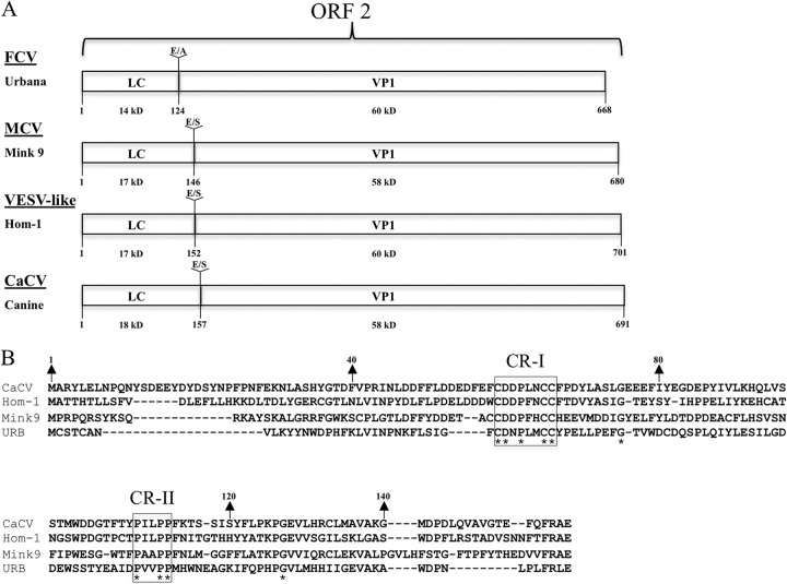
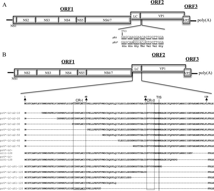
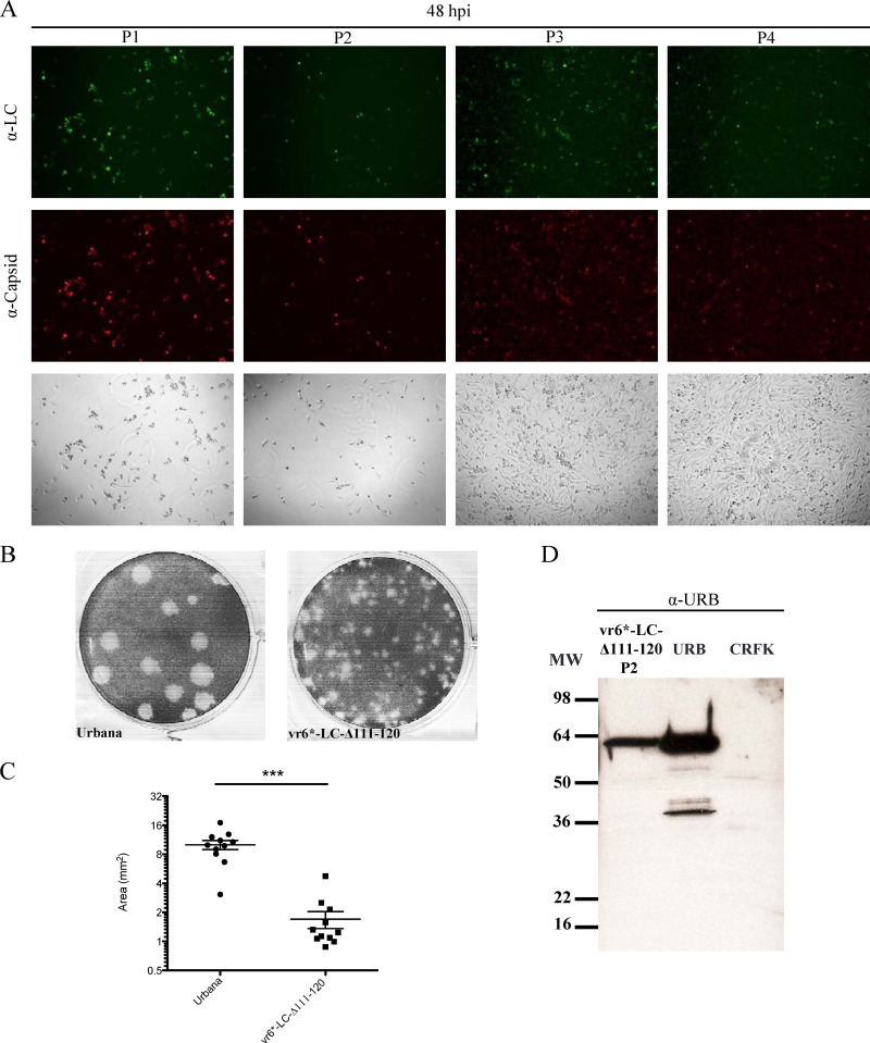
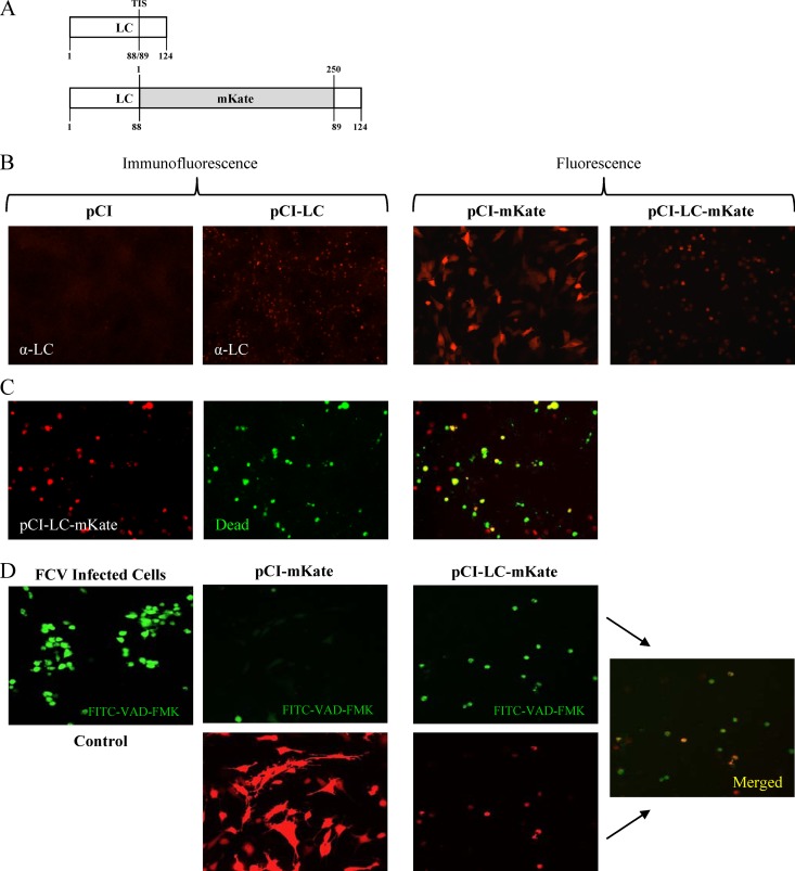
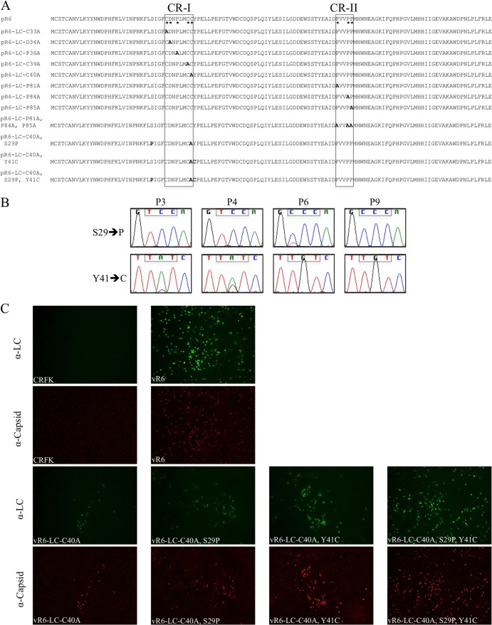
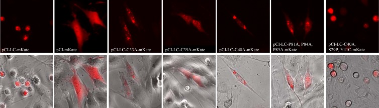
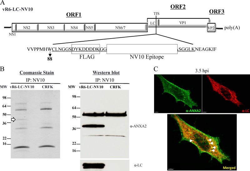
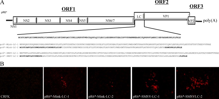
Open reading frame 2 (ORF2) of the feline calicivirus (FCV) genome encodes a capsid precursor that is posttranslationally processed to release the mature capsid protein (VP1) and a small protein of 124 amino acids, designated the leader of the capsid (LC). To investigate the role of the LC protein in the virus life cycle, mutations and deletions were introduced into the LC coding region of an infectious FCV cDNA clone. Three cysteine residues that are conserved among all vesivirus LC sequences were found to be critical for the recovery of FCV with a characteristic cytopathic effect in feline kidney cells. A cell-rounding phenotype associated with the transient expression of wild-type and mutagenized forms of the LC correlated with the cytopathic and growth properties of the corresponding engineered viruses. The host cellular protein annexin A2 was identified as a binding partner of the LC protein, consistent with a role for the LC in mediating host cell interactions that alter the integrity of the cell and enable virus spread.


Figures

Fig 1
Fig 1
Vesivirus LC sequences. (A) The organization of ORF2 from representative vesiviruses: FCV (Urbana), mink calicivirus 9 (MCV), VESV (Hom-1), and CaCV. Sequences of the dipeptide cleavage sites and their positions between the LC and VP1 are indicated. The calculated molecular mass of each protein is shown. (B) Amino acid alignment of the LC sequences of the four representative vesiviruses. Asterisks indicate residues conserved across all of the LC proteins that were analyzed. CR-I and CR-II refer to conserved regions.
Fig 2
Fig 2
Phylogenetic analysis of LC sequences. (A) Bayesian phylogenetic analysis of 88 LC nucleotide sequences showing the presence of four major clusters. (B) Amino acid p distances within and between the four clusters.
Fig 3
Fig 3
(A) Recombinant FL FCV clones with truncated LC sequences. The FCV genome is organized into three open reading frames (ORFs 1 to 3), with the ORF1 encoding nonstructural proteins 1 (NS1) through NS6/7. ORF2 encodes a capsid protein precursor that is cleaved by the viral proteinase (NS6/7) to yield LC and VP1. ORF3 encodes minor structural protein VP2. Two silent nucleotide changes (lowercase letters) were engineered into the FL FCV plasmid pR6 near the beginning of the VP1 coding sequence in ORF2 to create unique restriction enzyme site KpnI (boxed) in plasmid pR6* in order to facilitate cloning. Nucleotide triplets corresponding to VP1 codons are underlined, and the encoded amino acids are indicated below. (B) Plasmid constructions containing engineered deletions of the LC in the context of the FL genome. Amino acid numbers corresponding to the LC are indicated with arrows, regions conserved across all vesiviruses are boxed (CR-I and CR-II), and the transposon insertion site (TIS) is indicated with a line. The names of the FL clones refer to the LC amino acids that have been deleted.
Fig 4
Fig 4
Analysis of replication of vR6*-LC-Δ111–120. (A) Fluorescence microscopy analysis of LC and capsid expression in CRFK cells from a series of passages of vR6*-LC-Δ111-120 at the same time point. Bright-field images are shown to demonstrate the differences between the CPE observed at P1 and P2 versus P3 and P4. CRFK cells were stained 48 hpi with hyperimmune sera raised against either FCV virions or recombinant LC. (B) Representative image of plaque assays performed simultaneously, comparing wild-type FCV (Urbana) and P1 of vR6*-LC-Δ111-120. (C) Plaque size of viruses in panel B, measured using the software program GraphClick. The statistical significance of differences in size was calculated using an unpaired t test (P < 0.0001). (D) Western blot analysis of a cell lysate from P2 of vR6*-LC-Δ111-120. A cell lysate of Urbana-infected cells (URB) was included as a positive control, and an uninfected cell lysate (CRFK) was included as a negative control. MW, molecular weight.
Fig 5
Fig 5
Analysis of transient expression of the LC in CRFK cells. (A) Schematic representations of the wild-type LC protein and the recombinant LC-mKate fusion protein. The transposon insertion site (TIS) is indicated for the wild-type LC, and the location of the insertion of the red fluorescent protein mKate is shown. (B) Immunofluorescence and fluorescence analyses of CRFK cells transfected with the indicated plasmids. The LC was detected using hyperimmune sera, and expression of mKate and LC-mKate was visualized in live cells. (C) Representative images of CRFK cells transfected with pCI-LC-mKate and treated with the Live/Dead reagent (Invitrogen) that stains dead cells green. (D) Fluorescence microscopy of CRFK cells transfected with either pCI-mKate or pCI-LC-mKate and treated with FITC-VAD-FMK. FCV-infected cells were used as a positive control for apoptosis induction.
Fig 6
Fig 6
Alanine replacement of conserved residues in the LC in the FL FCV cDNA clone and analysis of virus capsid and LC expression. (A) FL plasmid constructions containing engineered alanine replacements of the conserved residues (*) from CR-I and CR-II. Additionally, three constructs that contain the C40A substitution and either one or both compensatory mutations (S29P and Y41C) are shown. The names of the FL clones refer to the LC amino acids that are replaced, their position according to the coding sequence of the LC, and the amino acid substitutions. (B) Chromatograms of LC codons (boxed nucleotides) at positions 29 and 41 corresponding to several different passages of vR6-LC-C40A. (C) Fluorescence microscopy analysis of LC and capsid expression in CRFK cells at P1 from the various FCV mutants. vR6-infected and mock-infected CRFK cells were included as positive and negative controls, respectively.
Fig 7
Fig 7
Alanine replacement of conserved residues in the LC in the pCI-LC-mKate clone and analysis of cell morphology in transient-expression experiments. Fluorescence microscopy analysis of CRFK cells transfected with LC-mKate, LC-mKate clones containing alanine replacements of conserved cysteines in the CR-I, and an LC-mKate-C40A clone that contained the two compensatory mutations required to suppress the defect in virus spread caused by the C40A substitution was performed. Fluorescent images are shown in the top row, and corresponding merged images (bright field and fluorescent) are shown below.
Fig 8
Fig 8
Coimmunoprecipitation of recombinant LC from infected CRFK cells. (A) A recombinant FL FCV clone in which two unique tags were introduced into the LC coding sequence at the TIS (between amino acids 88 and 89). The entire heterologous sequence inserted into the LC is underlined, and the two unique tags are boxed and labeled. FLAG refers to the commercially available FLAG tag, and the white box corresponds to the NV10 epitope of the Norwalk virus VP1 capsid protein, as described in Materials and Methods. (B) Coomassie blue stain and Western blot analyses of protein samples immunoprecipitated (IP) with the NV10 monoclonal antibody from vR6-LC-NV10- or mock-infected CRFK lysates separated by SDS-PAGE. The Coomassie blue stain revealed a band that was consistently observed at ∼36 kDa, as indicated by the white arrow. The Western blot was first probed with an anti-ANXA2 monoclonal antibody, stripped, and then reprobed with anti-LC hyperimmune sera. (C) Confocal images of CRFK cells infected with vR6 (MOI, 10). Cells were fixed 3.5 hpi. Immunofluorescence analysis was performed to detect ANXA2 (green) and LC (red). The bottom panel shows a merged image in which a yellow signal indicates colocalization of ANXA2 and LC. White arrows indicate sites of colocalization of ANXA2 and LC.
Fig 9
Fig 9
FL LC-chimeric FCV clones. (A) Recombinant FL FCV clones in which the FCV LC was replaced by either the entire or partial SMSV-5 Hom-1 or mink calicivirus 9 LC sequence. (B) Fluorescence microscopy analysis of capsid expression in CRFK cells at P1 from the various recombinant FCV mutants.

References

    1. Abente EJ, Sosnovtsev SV, Bok K, Green KY. 2010. Visualization of feline calicivirus replication in real-time with recombinant viruses engineered to express fluorescent reporter proteins. Virology 400:18–31 - PMC - PubMed
    1. Mitra T, Sosnovtsev SV, Green KY. 2004. Mutagenesis of tyrosine 24 in the VPg protein is lethal for feline calicivirus. J. Virol. 78:4931–4935 - PMC - PubMed
    1. Sosnovtsev S, Green KY. 1995. RNA transcripts derived from a cloned full-length copy of the feline calicivirus genome do not require VpG for infectivity. Virology 210:383–390 - PubMed
    1. Sosnovtsev SV, Belliot G, Chang KO, Onwudiwe O, Green KY. 2005. Feline calicivirus VP2 is essential for the production of infectious virions. J. Virol. 79:4012–4024 - PMC - PubMed
    1. Sosnovtsev SV, Sosnovtseva SA, Green KY. 1998. Cleavage of the feline calicivirus capsid precursor is mediated by a virus-encoded proteinase. J. Virol. 72:3051–3059 - PMC - PubMed

Publication types

LinkOut - more resources