. 2013 Mar;87(6):2994-3002.
doi: 10.1128/JVI.03017-12. Epub 2012 Dec 26.

Biophysical and ultrastructural characterization of adeno-associated virus capsid uncoating and genome release

Affiliations

Biophysical and ultrastructural characterization of adeno-associated virus capsid uncoating and genome release

Eric D Horowitz et al. J Virol. 2013 Mar.

Abstract

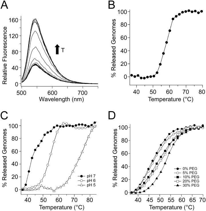
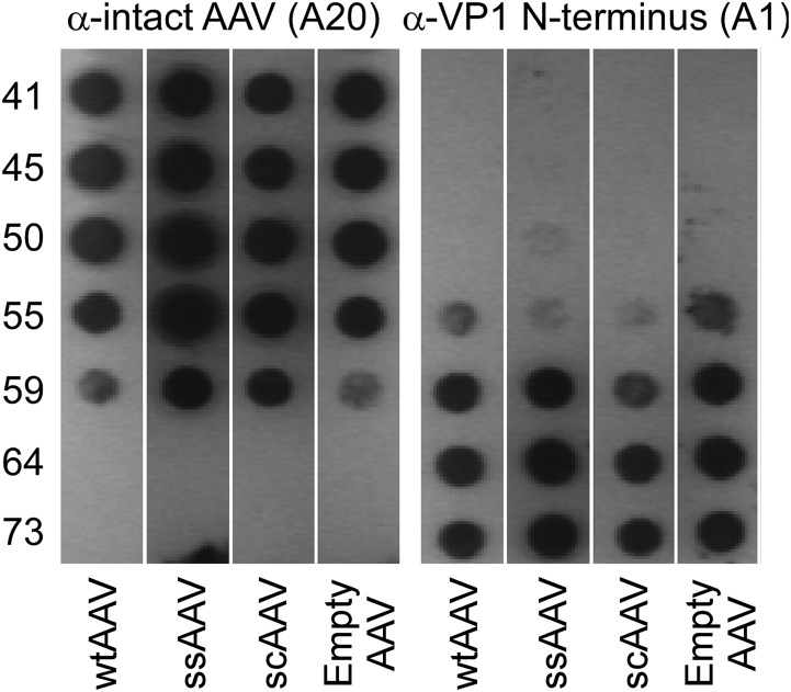
We describe biophysical and ultrastructural differences in genome release from adeno-associated virus (AAV) capsids packaging wild-type DNA, recombinant single-stranded DNA (ssDNA), or dimeric, self-complementary DNA (scDNA) genomes. Atomic force microscopy and electron microscopy (EM) revealed that AAV particles release packaged genomes and undergo marked changes in capsid morphology upon heating in physiological buffer (pH 7.2). When different AAV capsids packaging ss/scDNA varying in length from 72 to 123% of wild-type DNA (3.4 to 5.8 kb) were incrementally heated, the proportion of uncoated AAV capsids decreased with genome length as observed by EM. Genome release was further characterized by a fluorimetric assay, which demonstrated that acidic pH and high osmotic pressure suppress genome release from AAV particles. In addition, fluorimetric analysis corroborated an inverse correlation between packaged genome length and the temperature needed to induce uncoating. Surprisingly, scAAV vectors required significantly higher temperatures to uncoat than their ssDNA-packaging counterparts. However, externalization of VP1 N termini appears to be unaffected by packaged genome length or self-complementarity. Further analysis by tungsten-shadowing EM revealed striking differences in the morphologies of ssDNA and scDNA genomes upon release from intact capsids. Computational modeling and molecular dynamics simulations suggest that the unusual thermal stability of scAAV vectors might arise from partial base pairing and optimal organization of packaged scDNA. Our work further defines the biophysical mechanisms underlying adeno-associated virus uncoating and genome release.


Figures

Fig 1
Fig 1
Ultrastructural characterization of AAV capsid uncoating. (A) Atomic force microscopy images of scAAV (sc-CMV-GFP) vectors heated to 37°C and 65°C. Viral particles are observed as 25-nm spheres. After heating to 65°C, viral genomes are detected both associated with viral particles and as free DNA (arrowheads). (B) Observed morphologies of heat treated AAV. Genome-containing AAV particles are impermeable to uranyl acetate (i), while empty AAV particles that have released DNA exhibit a ring-like architecture (ii). Genome release resulted in diverse morphologies characterized by small gaps in the capsid wall (iii to v) as well as capsid fragments forming multimeric aggregates (vi to x). The scale bar represents 20 nm.
Fig 2
Fig 2
Effect of packaged genome length on AAV capsid uncoating. ssAAV (A) and scAAV (B) vectors were heated to different temperatures for 30 min prior to TEM imaging. Full AAV virions are viewed as 25-nm solid spheres, and empty virions are 25-nm donut-like structures. Differences in the relative amount of empty particles are apparent for different vector genome lengths. Genome lengths shown represent single-stranded DNA lengths (e.g., sc CMV-GFP is 2 × 2,058 bases + 3 × ITRs = 4,551 bases). Images are representative of 10 to 20 captured images. Scale bars are 50 nm.
Fig 3
Fig 3
Quantitative analysis of TEM images. Thermally induced uncoating efficiency was assessed for intact-genome-containing (full) AAV particles packaging ssDNA (A) and scDNA (B) genomes of different length. Vectors were heated to 37, 55, or 65°C and visualized with TEM. Averages are taken from 10 to 20 images with particle counts for each sample ranging from 395 to 5,317 total particles. In all cases the average and standard deviation at each temperature was determined using the percentage of full particles from each image. Smaller capsid fragments (<50% of the shell) were excluded from counting. Genome lengths shown represent single-stranded DNA lengths. All values at 55°C and 65°C were determined to be significantly different from those at 37°C (P < 0.001 by a two-tailed Student t test).
Fig 4
Fig 4
Fluorimetric detection of AAV genome release under different conditions. (A) In the presence of SYBR gold (25 μM), AAV capsids were heated from 37°C to 95°C. At the point of genome release, an increase in fluorescence is observed at 550 nm (excitation, 495 nm), arising from the interaction between SYBR gold and the AAV genome. (B) When plotted and normalized to pretransition and posttransition baselines, a relatively sharp transition is observed in fluorescence at the emission wavelength. (C and D) Encapsidation stability is increased with decreasing pH (C) as well as increasing osmotic pressure (D). All experiments were repeated in triplicate.
Fig 5
Fig 5
Fluorimetric analysis of AAV capsid uncoating as a function of genome length and self-complementarity. (A) Viral vectors were heated in 2°C increments and held for 5 min at each temperature prior to acquiring fluorescent signal. Three representative curves for different vector genome lengths are shown. Each vector was heated in triplicate along with a premelted control, which did not show a sharp transition. Curves were normalized to pre- and posttransition baselines to yield the percentage of released genomes. (B) Comparison of ssAAV, scAAV, and oversized ssAAV vectors shows an inverse linear correlation of Tm with vector genome size.
Fig 6
Fig 6
Exposure of the VP1 N termini is not dependent on genome length or self-complementarity. AAV capsids (5 × 10⁹ vg/well) containing either the wild type (wtAAV), single-stranded CMV-GFP (ssAAV), self-complementary CMV-GFP (scAAV), or no genome at all were heated in a PCR mixture for 30 min. The monoclonal antibodies A20 and A1 were then used to probe the AAV capsid integrity as well as the state of exposure of the buried VP1 N-terminal domain required for infection. No significant difference was observed between genomes with different sizes or self-complementarity.
Fig 7
Fig 7
Tungsten-shadowing EM of released genomes from ssCMV-GFP (A and C) and scCMV-GFP (B and D) vectors. Viral particles were heated to 65°C and then imaged using tungsten-shadowing EM. Grids showed a combination of AAV virions shown as spheres, loose DNA, and virion-associated DNA. Scale bars represent 50 nm. Insets are cartoon representations of the predicted secondary structure.
Fig 8
Fig 8
Computational modeling of internal capsid pressure and genome organization. (A) In molecular dynamics simulations, the stiffness of modeled ssDNA chains was increased step by step along the chain to simulate DNA base pairing in scAAV genomes. Pressure exerted on the capsid walls decreases until 50% of the chain has been converted, after which the capsid pressure increases. Error bars represent standard deviations (n = 5). Cartoon representations of predicted secondary structures of AAV genomes at different levels of base-pairing are shown above the curve. (B) Molecular dynamics simulation of progressive change in the organization of flexible ssDNA chains (cyan licorice) while being base paired into stiff dsDNA chains (red beads). Representative snapshots of chain organization within the model capsid at 0% (i), 25% (ii), 50% (iii), 75% (iv), and 100% (v) conversion of ssDNA to dsDNA are shown. dsDNA segments are seen to migrate to the periphery and adjacent to the inner capsid surface, where they may buffer the thermal motion of the flexible ssDNA chain and reduce the pressure exerted on the inner capsid walls during the initial stages of the conversion process. The development of an inner coil of dsDNA after 50% conversion may subsequently contribute to increasing capsid pressures.

References

    1. Bowles DE, Rabinowitz JE, Samulski RJ. 2006. The genus Dependovirus, p 15–24 In Kerr JR, Cotmore SF, Bloom ME, Linden RM, Parrish CR. (ed), Parvoviruses. Hodder Arnold Publishers, London, United Kingdom
    1. Samulski RJ, Srivastava A, Berns KI, Muzyczka N. 1983. Rescue of adeno-associated virus from recombinant plasmids: gene correction within the terminal repeats of AAV. Cell 33:135–143 - PubMed
    1. Xiao X, Xiao W, Li J, Samulski RJ. 1997. A novel 165-base-pair terminal repeat sequence is the sole cis requirement for the adeno-associated virus life cycle. J. Virol. 71:941–948 - PMC - PubMed
    1. Agbandje-McKenna M, Kleinschmidt J. 2011. AAV capsid structure and cell interactions. Methods Mol. Biol. 807:47–92 - PubMed
    1. Mingozzi F, High KA. 2011. Therapeutic in vivo gene transfer for genetic disease using AAV: progress and challenges. Nat. Rev. Genet. 12:341–355 - PubMed

Publication types

LinkOut - more resources