Unraveling of a neutralization mechanism by two human antibodies against conserved epitopes in the globular head of H5 hemagglutinin
- PMID: 23269809
- PMCID: PMC3592130
- DOI: 10.1128/JVI.01292-12
Unraveling of a neutralization mechanism by two human antibodies against conserved epitopes in the globular head of H5 hemagglutinin
Abstract
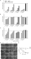
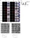
The rapid spread of highly pathogenic avian influenza (HPAI) H5N1 virus underscores the importance of effective antiviral treatment. Previously, we developed human monoclonal antibodies 65C6 and 100F4 that neutralize almost all (sub)clades of HPAI H5N1. The conserved 65C6 epitope was mapped to the globular head of HA. However, neither the 100F4 epitope nor the neutralization mechanism by these antibodies was known. In this study, we determined the 100F4 epitope and unraveled a neutralization mechanism by antibodies 65C6 and 100F4.
Figures


Similar articles
-
Divergent Requirement of Fc-Fcγ Receptor Interactions for In Vivo Protection against Influenza Viruses by Two Pan-H5 Hemagglutinin Antibodies.J Virol. 2017 May 12;91(11):e02065-16. doi: 10.1128/JVI.02065-16. Print 2017 Jun 1. J Virol. 2017. PMID: 28331095 Free PMC article.
-
A human antibody recognizing a conserved epitope of H5 hemagglutinin broadly neutralizes highly pathogenic avian influenza H5N1 viruses.J Virol. 2012 Mar;86(6):2978-89. doi: 10.1128/JVI.06665-11. Epub 2012 Jan 11. J Virol. 2012. PMID: 22238297 Free PMC article.
-
An antibody against a novel and conserved epitope in the hemagglutinin 1 subunit neutralizes numerous H5N1 influenza viruses.J Virol. 2010 Aug;84(16):8275-86. doi: 10.1128/JVI.02593-09. Epub 2010 Jun 2. J Virol. 2010. PMID: 20519402 Free PMC article.
-
Recent advances in "universal" influenza virus antibodies: the rise of a hidden trimeric interface in hemagglutinin globular head.Front Med. 2020 Apr;14(2):149-159. doi: 10.1007/s11684-020-0764-y. Epub 2020 Apr 1. Front Med. 2020. PMID: 32239416 Free PMC article. Review.
-
The antigenic architecture of the hemagglutinin of influenza H5N1 viruses.Mol Immunol. 2013 Dec;56(4):705-19. doi: 10.1016/j.molimm.2013.07.010. Epub 2013 Aug 7. Mol Immunol. 2013. PMID: 23933511 Review.
Cited by
-
Structural and functional definition of a vulnerable site on the hemagglutinin of highly pathogenic avian influenza A virus H5N1.J Biol Chem. 2019 Mar 22;294(12):4290-4303. doi: 10.1074/jbc.RA118.007008. Epub 2019 Feb 8. J Biol Chem. 2019. PMID: 30737282 Free PMC article.
-
Complementary recognition of the receptor-binding site of highly pathogenic H5N1 influenza viruses by two human neutralizing antibodies.J Biol Chem. 2018 Oct 19;293(42):16503-16517. doi: 10.1074/jbc.RA118.004604. Epub 2018 Aug 28. J Biol Chem. 2018. PMID: 30154240 Free PMC article.
-
B Cell Responses against Influenza Viruses: Short-Lived Humoral Immunity against a Life-Long Threat.Viruses. 2021 May 22;13(6):965. doi: 10.3390/v13060965. Viruses. 2021. PMID: 34067435 Free PMC article. Review.
-
Crimean-Congo haemorrhagic fever virus.Nat Rev Microbiol. 2023 Jul;21(7):463-477. doi: 10.1038/s41579-023-00871-9. Epub 2023 Mar 14. Nat Rev Microbiol. 2023. PMID: 36918725 Free PMC article. Review.
-
Association of poultry vaccination with interspecies transmission and molecular evolution of H5 subtype avian influenza virus.Sci Adv. 2025 Jan 24;11(4):eado9140. doi: 10.1126/sciadv.ado9140. Epub 2025 Jan 22. Sci Adv. 2025. PMID: 39841843 Free PMC article.
References
-
- Hu H, Voss J, Zhang G, Buchy P, Zuo T, Wang L, Wang F, Zhou F, Wang G, Tsai C, Calder L, Gamblin SJ, Zhang L, Deubel V, Zhou B, Skehel JJ, Zhou P. 2012. A human antibody recognizing a conserved epitope of H5 hemagglutinin broadly neutralizes highly pathogenic avian influenza H5N1 viruses. J. Virol. 86:2978–2989 - PMC - PubMed
-
- Tsai C, Caillet C, Hu H, Zhou F, Ding H, Zhang G, Zhou B, Wang S, Lu S, Buchy P, Deubel V, Vogel FR, Zhou P. 2009. Measurement of neutralizing antibody responses against H5N1 clades in immunized mice and ferrets using pseudotypes expressing influenza hemagglutinin and neuraminidase. Vaccine 27:6777–6790 - PMC - PubMed
-
- Caton AJ, Brownlee GG, Yewdell JW, Gerhard W. 1982. The antigenic structure of the influenza virus A/PR/8/34 hemagglutinin (H1 subtype). Cell 31:417–427 - PubMed
-
- Stray SJ, Pittman LB. 2012. Subtype- and antigenic site-specific differences in biophysical influences on evolution of influenza virus hemagglutinin. Virol. J. 9:91 doi:10.1186/1743-422X-9-91 - DOI - PMC - PubMed
Publication types
MeSH terms
Substances
LinkOut - more resources
Full Text Sources

