

doi: 10.12659/pjr.883628.
Bone marrow reconversion - imaging of physiological changes in bone marrow
Affiliations
- PMID: 23269936
- PMCID: PMC3529711
- DOI: 10.12659/pjr.883628
Item in Clipboard
Bone marrow reconversion - imaging of physiological changes in bone marrow
Pol J Radiol.
2012 Oct.
Abstract
Reconversion of bone marrow is a reverse process of natural replacement of red marrow by yellow marrow. The occurrence of reconversion can be misleading and challenging in interpretation of musculoskeletal system imaging. Changes of signal intensity in bone marrow are frequently observed in radiological routine and its diversity can cause a suspicion of pathologic findings. Therefore, the knowledge about distribution of red and yellow bone marrow depending on age, concomitant diseases and presentation of the patient are essential for MR image interpretation.
Keywords: bone marrow imaging; magnetic resonance; marrow reconversion.
Figures
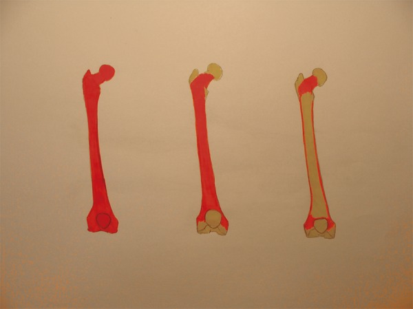
Graphic shows normal developmental transformation of bone marrow in the long bones (courtesy of L. Chudziak-Grędzicki).
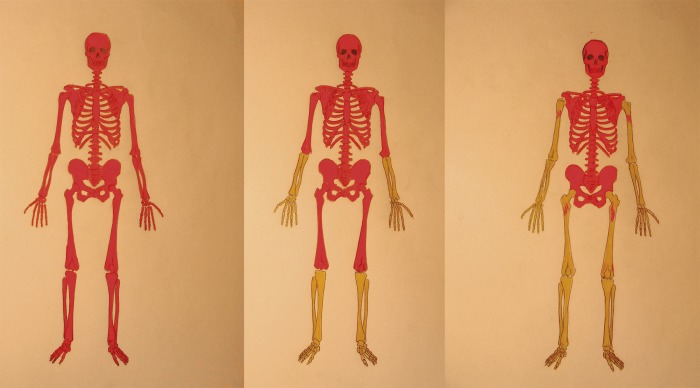
Graphic shows process of conversion of red-to-yellow bone marrow in the skeleton. On the left – red marrow fills all marrow cavities (at birth); the middle picture shows fatty conversion in the distal extremities (in childhood); the right picture shows progression of this process in the axial skeleton (courtesy of L. Chudziak-Grędzicki).
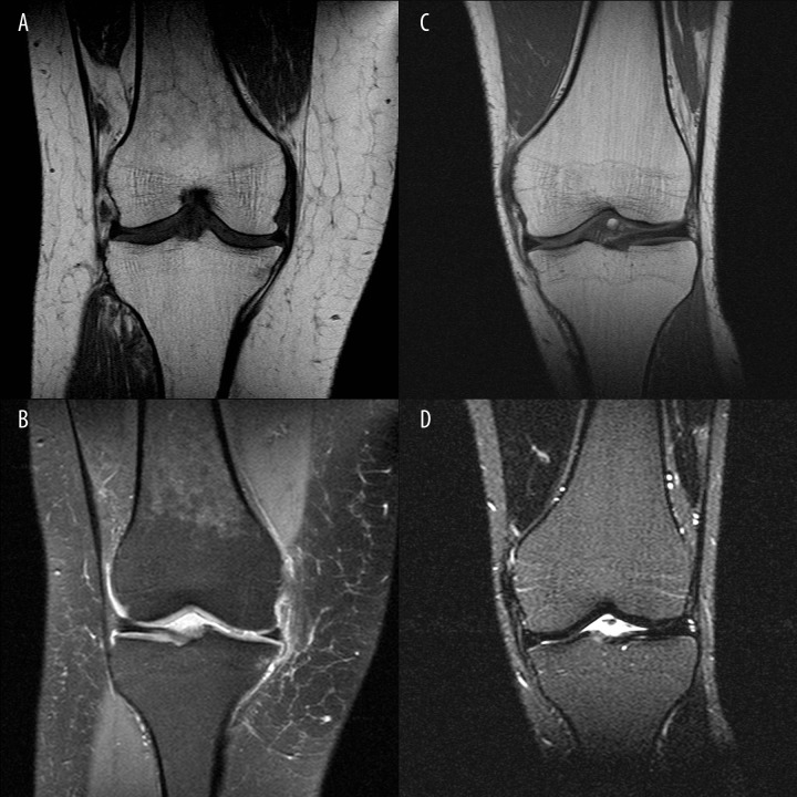
(A) Coronal T1-weighted image of the right knee joint in a 43-year old female patient reviewed due to chronic knee pain. In the distal femur there is patchy decrease in signal intensity, not extending beyond the line of growth plate. (B) PD fat saturation image. Clinical data from patient database CM Enel-Med S.A. (MAXIMA): obesity >25 BMI, smoking habit and type 2 diabetes treated with insulin. (C, D) Coronal T1-weighted and PD images of the knee joint in another 40-year old female without any known internal diseases show a typical for adults bone marrow picture in this area. The entire marrow cavity is filled with yellow bone marrow.
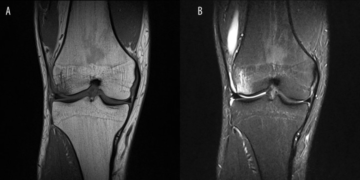
(A) Coronal T1-wieghted images of the knee joint in a 30yo patient after knee injury. Decreased signal intensity is visible in the marrow cavity (signal intensity of the muscles is lower than of marrow cavity), reaching but not extending beyond the growth plate. Patient is an amateur long-distance runner, with weekly routine over 70 km. (B) PD fat saturation images show decreased signal intensity of yellow bone marrow. The image also reveals the presence of bone marrow edema on the lateral femoral epicondyle. Signal intensity of edematous bone marrow is higher than in metaphyseal areas of reconversion.
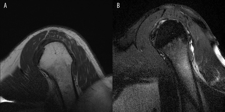
(A) Sagital oblique T1-weighted images of a 45 yo patient with shoulder pain shows areas of lower signal intensity in the metaphyseal area corresponding to bone marrow reconversion, which do not extend beyond the growth plate. Clinical data: smoking habit. (B) Sagital oblique PD fat saturation images in the same patient.
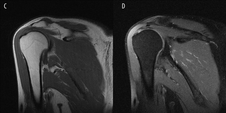
Coronal oblique T1-weighted and PD fat saturation images in a 40 yo patient – a picture typical for age.

Coronal T1-weighted images show areas of marrow reconversion in the proximal femora of a 33-year old female patient with microcytic anaemia treated with iron supplements.
References
-
- Vande Berg BC, Malghem J, Lecouvet FE, et al. Magnetic resonance imaging of normal bone marrow. Eur Radiol. 1998;8(8):1327–34. - PubMed
-
- Vande Berg BC, Malghem J, Lecouvet FE, et al. Magnetic resonance imaging of the bone marrow in hematological malignancies. Eur Radiol. 1998;8(8):1335–44. - PubMed
-
- Andrews CL. Evaluation of the Marrow Space in the Adult Hip. RadioGraphics. 2000;20:S27–42. - PubMed
-
- Ricci C, Cova M, Kang YS, et al. Normal Age-related Patterns of Cellular and Fatty Bone Marrow Distribution in the Axial Skeleton: MR Imaging Study. Radiology. 1990;177(1):83–88. - PubMed
-
- Laor T, Jaramillo D. MR imaging Insights into Skeletal Maturation: What Is Normal? Radiology. 2009;250:28–38. - PubMed
LinkOut - more resources
Full Text Sources