

doi: 10.5792/ksrr.2012.24.4.193.
Epub 2012 Nov 29.
Patellofemoral osteoarthritis
Affiliations
- PMID: 23269956
- PMCID: PMC3526755
- DOI: 10.5792/ksrr.2012.24.4.193
Item in Clipboard
Patellofemoral osteoarthritis
Knee Surg Relat Res.
2012 Dec.
Abstract
Patellofemoral arthritis is a fairly common disease, and it has been gaining interest with increasing number of studies due to its diverse treatment methods. Patellofemoral arthritis has a broad range of management options according to the characteristics of individual diseases. Identifying whether patellofemoral arthritis is the primary cause of knee pain and is compartment arthritis is necessary for establishing an adequate treatment method. Through investigation of the literature, the issues of recent knowledge of femoropatella arthritis and the diagnosis and treatment of which were studied.
Keywords: Diagnosis; Patellofemoral arthritis; Treatment.
Figures
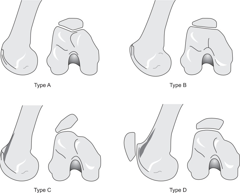
Dejour's classification of trochlear dysplasia. Type A: crossing sign (flat or convex trochlea), Type B: crossing sign and supratrochlear spur, Type C: crossing sign and double contour, Type D: crossing sign. Supratrochlear spur, double contour, and sharp step-off of the trochlea.
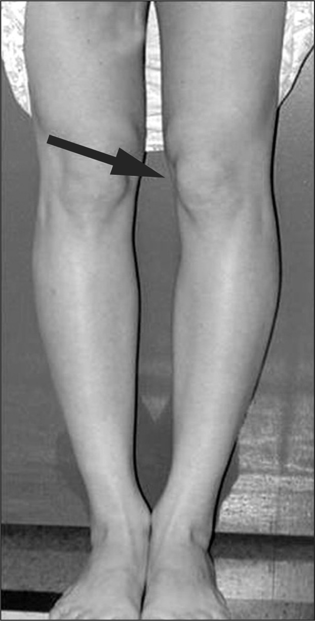
The points inward (arrow) are squinting patella. This finding is associated with femoral antevesion.
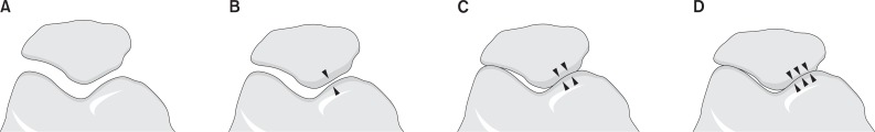
Merchant staged the severity of the disease based on the 45° skyline view. (A) Stage 1: mild with more than 3 mm of joint space. (B) Stage 2: moderate with less than 3 mm of joint space but no bony contact. (C) Stage 3: severe with bony surfaces in contact over less than one quarter of the joint surface. (D) Stage 4: very severe with bony contact throughout the entire joint surface.
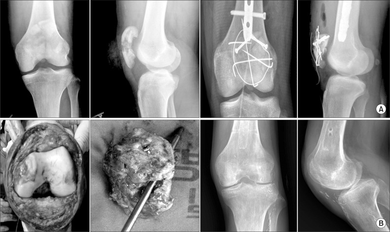
(A) Simple radiographs of a patient diagnosed with patellofemoral arthritis after patellar fracture. (B) Intraoperative gross photos and postoperative radiographs after patellectomy.
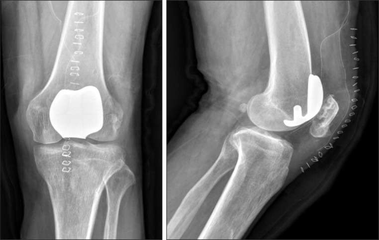
Postoperative simple radiographs after patellofemoral replacement in a patient diagnosed with patellar malunion.
References
-
- Davies AP, Vince AS, Shepstone L, Donell ST, Glasgow MM. The radiologic prevalence of patellofemoral osteoarthritis. Clin Orthop Relat Res. 2002;(402):206–212. - PubMed
-
- Noble J, Hamblen DL. The pathology of the degenerate meniscus lesion. J Bone Joint Surg Br. 1975;57:180–186. - PubMed
-
- Arendt EA, Fithian DC, Cohen E. Current concepts of lateral patella dislocation. Clin Sports Med. 2002;21:499–519. - PubMed
-
- Saleh KJ, Arendt EA, Eldridge J, Fulkerson JP, Minas T, Mulhall KJ. Symposium Operative treatment of patellofemoral arthritis. J Bone Joint Surg Am. 2005;87:659–671. - PubMed
-
- Mow VC, Kuei SC, Lai WM, Armstrong CG. Biphasic creep and stress relaxation of articular cartilage in compression? Theory and experiments. J Biomech Eng. 1980;102:73–84. - PubMed
LinkOut - more resources
Full Text Sources
Medical