

Role of stem/progenitor cells in reparative disorders
- PMID: 23270300
- PMCID: PMC3541267
- DOI: 10.1186/1755-1536-5-20
Role of stem/progenitor cells in reparative disorders
Abstract
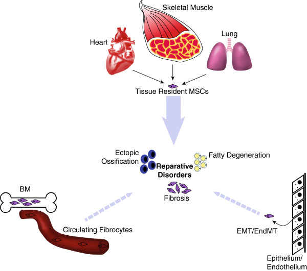
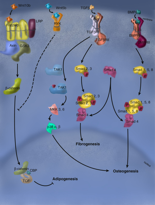
Adult stem cells are activated to proliferate and differentiate during normal tissue homeostasis as well as in disease states and injury. This activation is a vital component in the restoration of function to damaged tissue via either complete or partial regeneration. When regeneration does not fully occur, reparative processes involving an overproduction of stromal components ensure the continuity of tissue at the expense of its normal structure and function, resulting in a "reparative disorder". Adult stem cells from multiple organs have been identified as being involved in this process and their role in tissue repair is being investigated. Evidence for the participation of mesenchymal stromal cells (MSCs) in the tissue repair process across multiple tissues is overwhelming and their role in reparative disorders is clearly demonstrated, as is the involvement of a number of specific signaling pathways. Transforming growth factor beta, bone morphogenic protein and Wnt pathways interact to form a complex signaling network that is critical in regulating the fate choices of both stromal and tissue-specific resident stem cells (TSCs), determining whether functional regeneration or the formation of scar tissue follows an injury. A growing understanding of both TSCs, MSCs and the complex cascade of signals regulating both cell populations have, therefore, emerged as potential therapeutic targets to treat reparative disorders. This review focuses on recent advances on the role of these cells in skeletal muscle, heart and lung tissues.
Figures
Similar articles
-
New insights on the reparative cells in bone regeneration and repair.Biol Rev Camb Philos Soc. 2021 Apr;96(2):357-375. doi: 10.1111/brv.12659. Epub 2020 Oct 13. Biol Rev Camb Philos Soc. 2021. PMID: 33051970
-
Mesenchymal Stromal Cells and Tissue-Specific Progenitor Cells: Their Role in Tissue Homeostasis.Stem Cells Int. 2016;2016:4285215. doi: 10.1155/2016/4285215. Epub 2015 Dec 28. Stem Cells Int. 2016. PMID: 26823669 Free PMC article. Review.
-
Embryonic development and the principles of tissue engineering.Novartis Found Symp. 2003;249:17-25; discussion 25-33, 170-4, 239-41. Novartis Found Symp. 2003. PMID: 12708647 Review.
-
Role of Wnt/β-Catenin Signaling in Epithelial Differentiation of Lung Resident Mesenchymal Stem Cells.J Cell Biochem. 2015 Aug;116(8):1532-9. doi: 10.1002/jcb.25069. J Cell Biochem. 2015. PMID: 25546504
-
Immunomodulatory capacity of the local mesenchymal stem cells transplantation after severe skeletal muscle injury in female rats.Immunopharmacol Immunotoxicol. 2016 Dec;38(6):414-422. doi: 10.1080/08923973.2016.1222617. Epub 2016 Aug 25. Immunopharmacol Immunotoxicol. 2016. PMID: 27560658
Cited by
-
Mesenchymal stromal cell secreted sphingosine 1-phosphate (S1P) exerts a stimulatory effect on skeletal myoblast proliferation.PLoS One. 2014 Sep 29;9(9):e108662. doi: 10.1371/journal.pone.0108662. eCollection 2014. PLoS One. 2014. PMID: 25264785 Free PMC article.
-
Surgical Excision of Heterotopic Ossification Leads to Re-Emergence of Mesenchymal Stem Cell Populations Responsible for Recurrence.Stem Cells Transl Med. 2017 Mar;6(3):799-806. doi: 10.5966/sctm.2015-0365. Epub 2016 Oct 5. Stem Cells Transl Med. 2017. PMID: 28297577 Free PMC article.
-
Maintenance of vascular integrity by pericytes is essential for normal kidney function.Am J Physiol Renal Physiol. 2016 Dec 1;311(6):F1230-F1242. doi: 10.1152/ajprenal.00030.2016. Epub 2016 Jun 22. Am J Physiol Renal Physiol. 2016. PMID: 27335372 Free PMC article.
-
Plasticity of the Muscle Stem Cell Microenvironment.Adv Exp Med Biol. 2017;1041:141-169. doi: 10.1007/978-3-319-69194-7_8. Adv Exp Med Biol. 2017. PMID: 29204832 Free PMC article. Review.
-
Stem cells in dermatology.An Bras Dermatol. 2014 Mar-Apr;89(2):286-91. doi: 10.1590/abd1806-4841.20142530. An Bras Dermatol. 2014. PMID: 24770506 Free PMC article. Review.
References
LinkOut - more resources
Full Text Sources
