Review
. 2013 Feb;25(1):47-56.
doi: 10.1016/j.ceb.2012.10.018. Epub 2012 Dec 25.

The expanding significance of keratin intermediate filaments in normal and diseased epithelia

Affiliations
Review

The expanding significance of keratin intermediate filaments in normal and diseased epithelia

Xiaoou Pan et al. Curr Opin Cell Biol. 2013 Feb.

Abstract

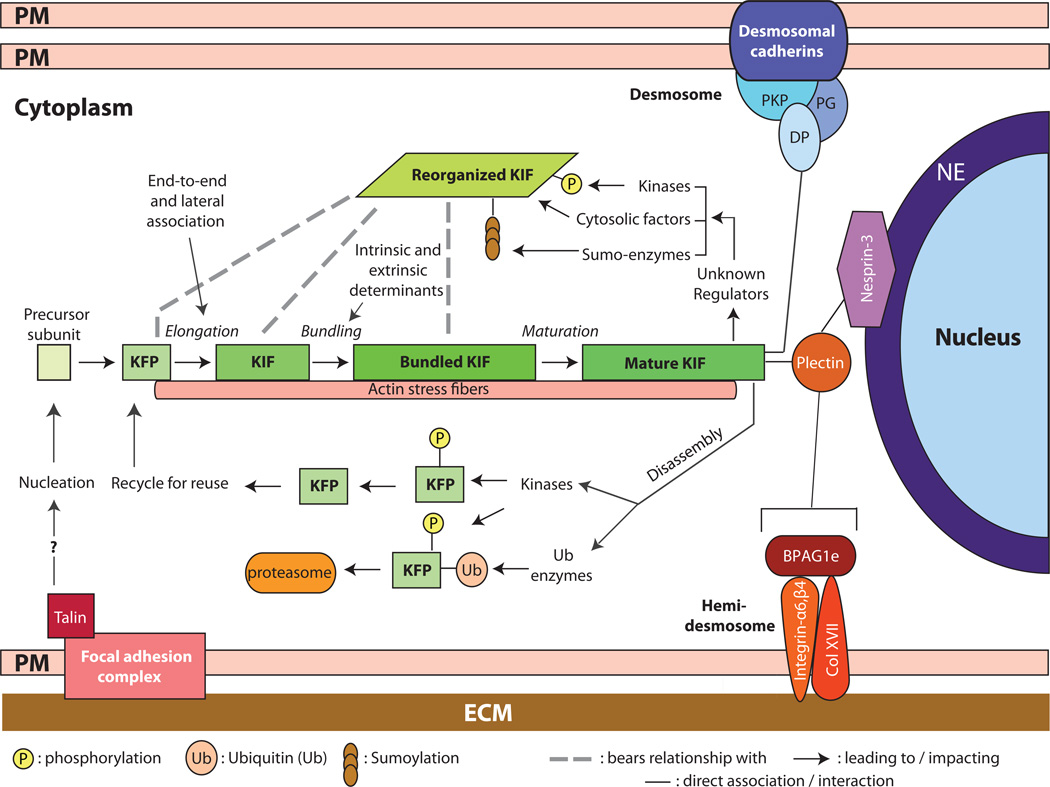
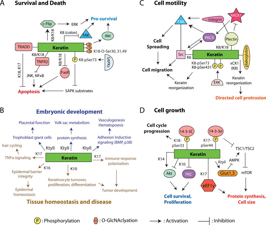
Intermediate filaments are assembled from a diverse group of evolutionary conserved proteins and are specified in a tissue-dependent, cell type-dependent, and context-dependent fashion in the body. Genetic mutations in intermediate filament proteins account for a large number of diseases, ranging from skin fragility conditions to cardiomyopathies and premature aging. Keratins, the epithelial-specific intermediate filaments, are now recognized as multi-faceted effectors in their native context. In this review, we emphasize the recent progress made in defining the role of keratins towards the regulation of cytoarchitecture, cell growth and proliferation, apoptosis, and cell motility during embryonic development, in normal adult tissues, and in select diseases such as cancer.


Figures

Figure 1
Figure 1
Assembly, organization, and regulation of keratin intermediate filaments (KIFs). Live imaging studies in epithelial cells in culture show that keratin filament assembly is initiated at the periphery of the cell, near focal adhesions, and that newly formed filaments and their maturation into an organized network takes place in the context of a continuous centripetal flow with disassembly and turnover steps taking place near the nuclear envelope. The resulting “keratin cycle” is highly dependent on interactions with F-actin, with additional proteins, and on several types of post-translational modifications including phosphorylation, ubiquitination, sumoylation and (though not shown here) O-glycosylation. Most likely, the biological context dictates the rate of flow through this cycle. The figure also conveys that KIFs are attached at the surface of the nucleus (via a plectin/Nesprin-3 complex), at desmosome cell-cell adhesion sites, (via desmoplakin (DP), among other proteins), and at hemidemosome cell-matrix adhesions (via plectin and BPAG1e). Not shown here are the interactions with F-actin and microtubules. The structural support role of KIFs depends upon their organization as a crosslinked network that is fully integrated with other structural elements within and between epithelial cells. NE: nuclear envelope; PM: plasma membrane; ECM: extracellular matrix; DP: desmoplakin; PKP: plakophilin; KFP: keratin filament precursor; KIF: keratin intermediate filament.
Figure 2
Figure 2
Examples of cellular and physiological processes regulated by keratin proteins. A) Cell survival and cell death. B) Embryonic development (top; blue lettering) and tissue homeostasis and disease (bottom; brown lettering). C) Cell motility and related processes. D) Cell growth. In all cases, lines connecting keratins and the molecule(s) of interest convey a physical interaction. Abbreviations: Akt, Protein kinase B; AMPK, AMP-activated protein kinase; eEF1γ, Eukaryotic translation elongation factor 1 gamma; ERK, Extracellular signal-regulated kinase; FAK, Focaladhesion kinase-1; FasR, Fas-ligand receptor; Glut1, 3, glucose transporters 1, 3; K, mammalian keratins; KtyII, Type II mammalian keratins; mTOR, Mammalian target of rapamycin; PKC, protein kinase C; RACK1, Receptor for activated protein kinase C; SAPK, stress activated protein kinases; Src, Src kinase; TNFR2, Tumor necrosis factor receptor 2; TRADD, Tumor necrosis factor receptor type I-associated Death domain protein; TSC1/2, Tuberous Sclerosis Complex (Hamartin/Tuberin); xCK, Xenopus cytokeratin (ortholog to mammalian K18).

References

    1. Schweizer J, Bowden PE, Coulombe PA, Langbein L, Lane EB, Magin TM, Maltais L, Omary MB, Parry DA, Rogers MA, Wright MW. New consensus nomenclature for mammalian keratins. J Cell Biol. 2006;174:169–174. - PMC - PubMed
    1. Fuchs E. Keratins and the skin. Ann. Rev. Cell Dev. Biol. 1995;11:123–153. - PubMed
    1. Moll R, Franke WW, Schiller DL, Geiger B, Krepler R. The catalog of human cytokeratins: patterns of expression in normal epithelia, tumors and cultured cells. Cell. 1982;31:11–24. - PubMed
    1. Toivola DM, Strnad P, Habtezion A, Omary MB. Intermediate filaments take the heat as stress proteins. Trends Cell Biol. 2010;20:79–91. - PMC - PubMed
    1. Kim S, Coulombe PA. Intermediate filament scaffolds fulfill mechanical, organizational, and signaling functions in the cytoplasm. Genes Dev. 2007;21:1581–1597. - PubMed

Publication types

LinkOut - more resources