. 2012:3:1329.
doi: 10.1038/ncomms2339.

Extracellular Ca2+ is a danger signal activating the NLRP3 inflammasome through G protein-coupled calcium sensing receptors

Affiliations
Free PMC article

Extracellular Ca2+ is a danger signal activating the NLRP3 inflammasome through G protein-coupled calcium sensing receptors

Manuela Rossol et al. Nat Commun. 2012.
Free PMC article

Abstract

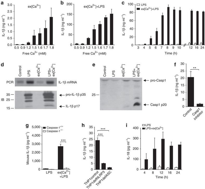
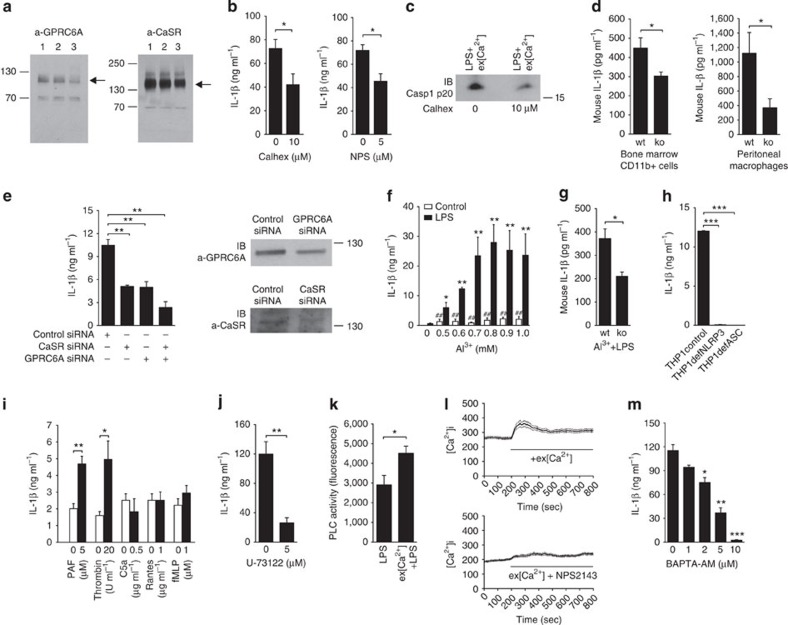
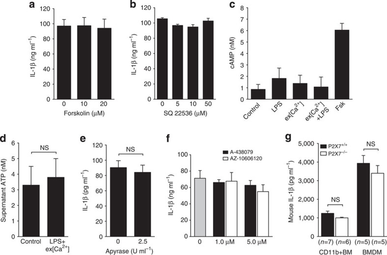
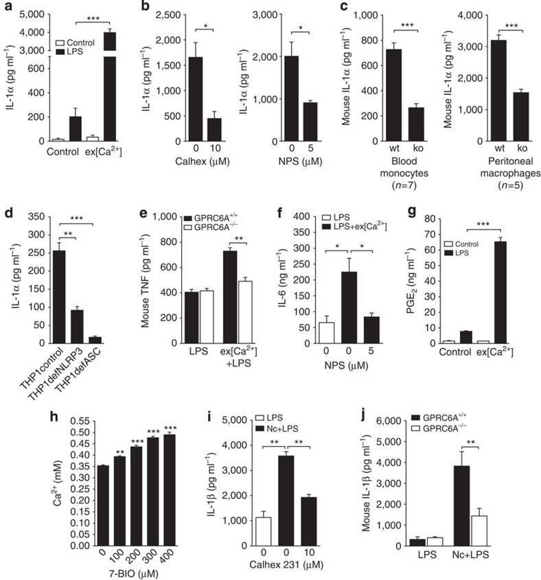
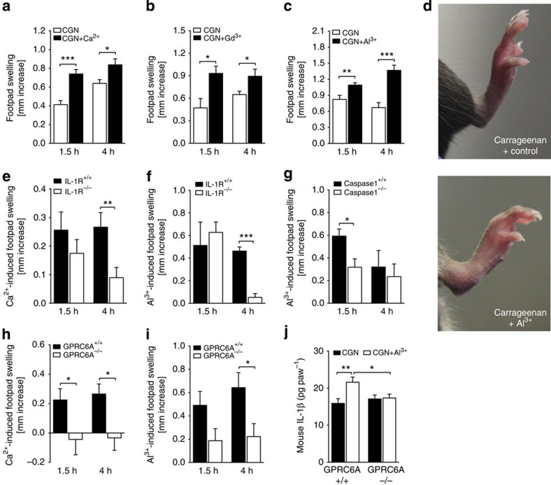
Activation of the NLRP3 inflammasome enables monocytes and macrophages to release high levels of interleukin-1β during inflammatory responses. Concentrations of extracellular calcium can increase at sites of infection, inflammation or cell activation. Here we show that increased extracellular calcium activates the NLRP3 inflammasome via stimulation of G protein-coupled calcium sensing receptors. Activation is mediated by signalling through the calcium-sensing receptor and GPRC6A via the phosphatidyl inositol/Ca(2+) pathway. The resulting increase in the intracellular calcium concentration triggers inflammasome assembly and Caspase-1 activation. We identified necrotic cells as one source for excess extracellular calcium triggering this activation. In vivo, increased calcium concentrations can amplify the inflammatory response in the mouse model of carrageenan-induced footpad swelling, and this effect was inhibited in GPRC6A(-/-) mice. Our results demonstrate that G-protein-coupled receptors can activate the inflammasome, and indicate that increased extracellular calcium has a role as a danger signal and amplifier of inflammation.


Figures

Figure 1
Figure 1. Extracellular calcium induces monocyte IL-1β release by activating the inflammasome.
(a,b) IL-1β release of CD14+ monocytes in response to increasing ex[Ca2+] alone (a) or to increasing ex[Ca2+] plus LPS (b) after 16 h of stimulation (n=3). (c) time course of IL-1β release of CD14+ monocytes after stimulation with LPS alone or with increased ex[Ca2+] (1.7 mM) plus LPS (n=3). (d) PCR (upper panel) and immunoblot (IB, lower panel) for IL-1β in unstimulated monocytes (control) and in monocytes stimulated with LPS, with increased ex[Ca2+] concentration (1.7 mM), or both, after 16 h of culture. Specific bands for IL-1β mRNA, pro-IL-1β p35 and IL-1β p17 as indicated. One representative experiment out of three is shown. (e) IB analysis of Caspase-1, which shows the activation of Caspase-1 in supernatants harvested from unstimulated monocytes (control) and in monocytes stimulated with LPS, with increased ex[Ca2+] concentration (1.7 mM), or with both, after 16 h of culture. Specific bands for pro-Caspase-1 (pro-Casp1) and Caspase-1 (Casp1 p20) are indicated, shown is one representative blot out of three experiments performed. (f) IL-1β secretion of monocytes stimulated with ex[Ca2+] plus LPS in the absence and presence of the caspase-1 inhibitor Z-WEHD-FMK (5 μM, n=3). (g) mIL-1β secretion of monocytes from Caspase-1+/+ and Caspase-1−/− NOD mice stimulated with LPS or with ex[Ca2+] plus LPS (n=3). (h) IL-1β secretion of THP-1 cells (control), NLRP3-deficient THP-1 cells (THP1defNLRP3) and ASC-deficient THP-1 cells (THP1defASC) stimulated with increased ex[Ca2+] concentration (1.7 mM) and LPS, after 16 h of culture (n=3). (i) Time course of IL-18 release of CD14+ monocytes after stimulation with LPS alone or with increased ex[Ca2+] (1.7 mM) plus LPS (n=3). In all experiments, cytokine concentrations were determined in the supernatant by enzyme-linked immunosorbent assay. All bars show mean±s.e.m. Statistical analysis was performed using the two-tailed Student’s t-test; **P<0.01 and ***P<0.001.
Figure 2
Figure 2. Extracellular calcium activates the inflammasome via CaSR and GPRC6A.
(a) Immunoblot (IB) analysis demonstrating the expression of GPRC6A (left panel) and CaSR (right) in human monocytes. In both blots, lanes 1, 2 and 3 represent three different healthy donors. (b) Influence of Calhex231 and NPS2143 on the IL-1β secretion of monocytes stimulated with increased ex[Ca2+]+LPS (n=3). (c) IB analysis showing the active form of caspase-1 (p20) in supernatants collected from monocytes stimulated with increased ex[Ca2+]+LPS in the absence or presence of Calhex231 after 16 h of culture. One representative blot out of three experiments performed is shown. (d) Ex[Ca2+]-induced IL-1β secretion of CD11b+ mononuclear cells isolated from bone marrow and of peritoneal macrophages from GPRC6A+/+ (wt) and GPRC6A−/− (ko) mice (n=5). (e) Influence of CaSR and GPRC6A inactivation by transfection with siRNA on IL-1β secretion of THP-1 cells in response to ex[Ca2+]+LPS (n=3). IBs show inhibition of protein expression of GPRC6A and CaSR in cells transfected with the respective siRNA compared with control siRNA. (f) IL-1β secretion of monocytes in response to increasing concentration of Al3+ with or without LPS (n=3). (g) IL-1β secretion of peritoneal macrophages from GPRC6A+/+ (wt) and GPRC6A−/− (ko) mice in response to 0.9mM Al3+ plus LPS (n=3). (h) IL-1β secretion of THP-1 cells (control), NLRP3-deficient THP-1 cells (THP1defNLRP3) and ASC-deficient THP-1 cells (THP1defASC) stimulated with 0.9 mM Al3+ plus LPS, for 16 h (n=3). (i) IL-1β release of monocytes stimulated with different GPCR ligands at the indicated concentration alone (white bars) or with GPCR ligands +LPS (black bars) for 16 h. (j) IL-1β release from monocytes stimulated with ex[Ca2+]+LPS in the presence of dimethyl sulfoxide (control) or with the phosphoinositide-specific PLC inhibitor U-73122 (5 μM) in dimethyl sulfoxide (n=3). (k) PLC activity in monocytes stimulated for 16 h with LPS alone or with ex[Ca2+]+LPS (n=5). (l) For [Ca2+]i imaging, monocytes were loaded with the Ca2+-sensitive indicator dye Fura-2-AM and subsequently stimulated with ex[Ca2+] (1.7 mM). The increase in [Ca2+]i was inhibited by NPS2143. One representative experiment out of three is shown. (m) IL-1β release from monocytes stimulated with ex[Ca2+]+LPS in the presence of increasing concentrations of BAPTA-AM (n=3). *P<0.05, **P<0.01 and ***P<0.001, always in comparison with the negative control. Statistical analysis was performed using the two-tailed Student's t-test. All error bars show mean±s.e.m.
Figure 3
Figure 3. The cAMP pathway and extracellular ATP is not involved in ex[Ca2+]-induced inflammasome activation.
(a,b) The indicated concentrations of forskolin (a) and of the adenylyl cyclase inhibitor SQ22536 (b) were added to monocyte cultures, and ex[Ca2+] (1.7 mM)-induced IL-1β production was determined (n=3). (c) Intracellular cAMP concentrations in response to ex[Ca2+] (1.7 mM) +LPS (n=3, mean±s.e.m.). Forskolin (Fsk) was used as positive control. All bars show mean±s.e.m. Statistical analysis was performed using a two-tailed Student’s t-test, and differences are not statistically significant. (a) ATP concentration in the supernatant of monocytes stimulated with ex[Ca2+] (1.7 mM) +LPS and of unstimulated monocytes (control). (b,c) IL-1β secretion of monocytes stimulated with ex[Ca2+] (1.7 mM) +LPS in the presence of the P2X7 inhibitors A-438079 and AZ-10606120 (b) or apyrase (c) at the indicated concentrations. (d) The ex[Ca2+] +LPS induced IL-1β secretion of CD11b+mononuclear cells isolated from bone marrow and bone marrow-derived macrophages from P2X7+/+ and P2X7−/− mice. (ad) Experiments were performed in triplicates. All bars show mean±s.e.m. Statistical analysis was performed using the two-tailed Student’s t-test, and differences are not statistically significant.
Figure 4
Figure 4. Extracellular calcium induces several proinflammatory cytokines and is released from necrotic cells.
(a) IL-1α release from unprimed (white bars) and LPS-primed (black bars) monocytes in response to stimulation with ex[Ca2+] (n=3). (b) Influence of the specific inhibitors Calhex231 and NPS2143 on IL-1α secretion of monocytes stimulated with increased ex[Ca2+]+LPS (n=3). (c) ex[Ca2+]-induced IL-1α secretion of CD11b+ mononuclear cells isolated from peripheral blood and of peritoneal macrophages from GPRC6A+/+ (wt) and GPRC6A−/− (ko) mice. (d) IL-1α secretion of THP-1 cells (control), NLRP3-deficient THP-1 cells (THP1defNLRP3) and ASC-deficient THP-1 cells (THP1defASC) stimulated with increased ex[Ca2+] concentration and LPS, after 16 h of culture (n=3). (e) Secretion of TNF from LPS-stimulated peritoneal macrophages or from macrophages stimulated with ex[Ca2+]+LPS from GPRC6A+/+ (wt) or GPRC6A−/− (ko) mice (n=3). (f) Influence of the specific inhibitor NPS2143 on IL-6 secretion of monocytes stimulated with increased ex[Ca2+]+LPS (n=3). (g) Prostaglandin E2 (PGE2) release from unprimed (white bars) and LPS-primed (black bars) monocytes in response to stimulation with ex[Ca2+] (n=3). (h) Influence of 7-BIO-induced monocyte necrosis on the Ca2+ concentration in the supernatant (n=3). (i) IL-1β release from LPS-primed CD14+ monocytes cultured alone (white bars) or co-cultured with necrotic autologous CD4+T cells (black bars) in the absence or presence of the specific inhibitor Calhex231 (n=3). (j) mIL-1β secretion of LPS-primed monocytes from GPRC6A+/+ (wt) and GPRC6A−/− (ko) mice cultured alone (LPS) or co-cultured with necrotic autologous CD4+ T cells (Nc+LPS, n=9). All bars show mean±s.e.m. Statistical analysis was performed using the two-tailed Student’s t-test. *P<0.05, **P<0.01 and ***P<0.001.
Figure 5
Figure 5. CaSR and GPRC6A are involved in inflammatory responses in vivo.
(ac) Carrageenan (CGN)-induced footpad swelling in C57BL/6 mice with (black bars) or without (white bars) co-injection of Ca2+ (n=28) (a), Al3+ (n=10) (c,d) or Gd3+ (n=6) (b). Bars depict the increase in paw thickness after 1.5 and 4 h compared with baseline. (d) Representative images of CGN-induced footpad swelling after 4 h with (right) or without (left) co-injection of Al3+. (e,f) Ca2+-induced (n=20; e) and Al3+-induced (n=4; f) increase in footpad swelling compared with Carrageenan alone in IL-1R+/+ and IL-1R−/− mice after 1.5 and 4 h. (g) Al3+-induced (n=4) increase in footpad swelling compared with Carrageenan alone in Caspase-1+/+ and Caspase-1−/− mice after 1.5 and 4 h. (h,i) Ca2+-induced (n=10; h) and Al3+-induced (n=12; i) increase in footpad swelling compared with Carrageenan alone in GPRC6A+/+ and GPRC6A−/− mice after 1.5 and 4 h. (j) IL-1β amount in exudats from Carageenan (CGN) or CGN plus Al3+-treated paws from GPRC6A+/+ (n=15) and GPRC6A−/− (n=17) mice. All data are expressed as mean±s.e.m. Statistical analysis was performed using a two-tailed Student’s t-test, *P<0.05, **P<0.01 and ***P<0.001.

Similar articles

Cited by

References

    1. Martinon F., Burns K., Tschopp J. The inflammasome: a molecular platform triggering activation of inflammatory caspases and processing of proIL-beta. Mol. Cell. 10, 417–426 (2002). - PubMed
    1. Chen G. Y., Nuñez G. Sterile inflammation: sensing and reacting to damage. Nat. Rev. Immunol. 10, 826–837 (2010). - PMC - PubMed
    1. Robertson W. G., Marshall R. W. Ionized calcium in body fluids. Crit. Rev. Clin. Lab. Sci. 15, 85–125 (1981). - PubMed
    1. Janle E. M., Sojka J. E. Use of ultrafiltration probes in sheep to collect interstitial fluid for measurement of calcium and magnesium. Contemp. Top. Lab. Anim. Sci. 39, 47–50 (2000). - PubMed
    1. Menkin In Biochemical Mechanisms in Inflammation (ed. Thomas) Springfield, IL (1958).

Publication types

MeSH terms