. 2013 Feb 8;288(6):3739-52.
doi: 10.1074/jbc.M112.426205. Epub 2012 Dec 27.

Oxidation-sensing regulator AbfR regulates oxidative stress responses, bacterial aggregation, and biofilm formation in Staphylococcus epidermidis

Affiliations

Oxidation-sensing regulator AbfR regulates oxidative stress responses, bacterial aggregation, and biofilm formation in Staphylococcus epidermidis

Xing Liu et al. J Biol Chem. .

Abstract

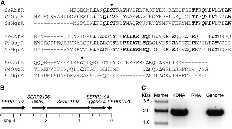
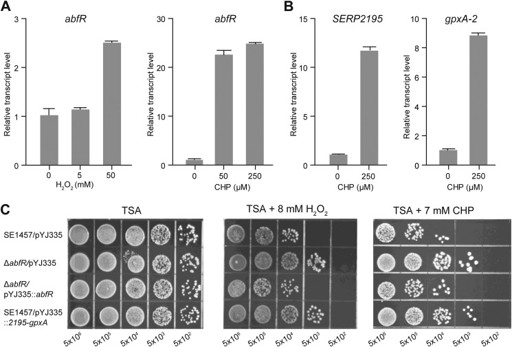
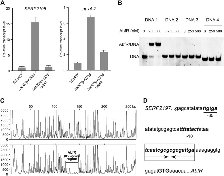
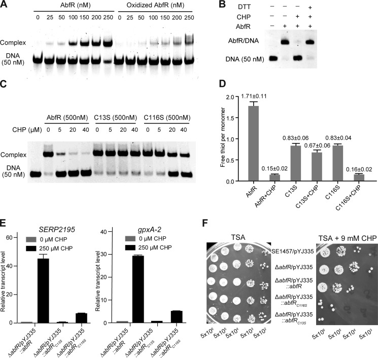
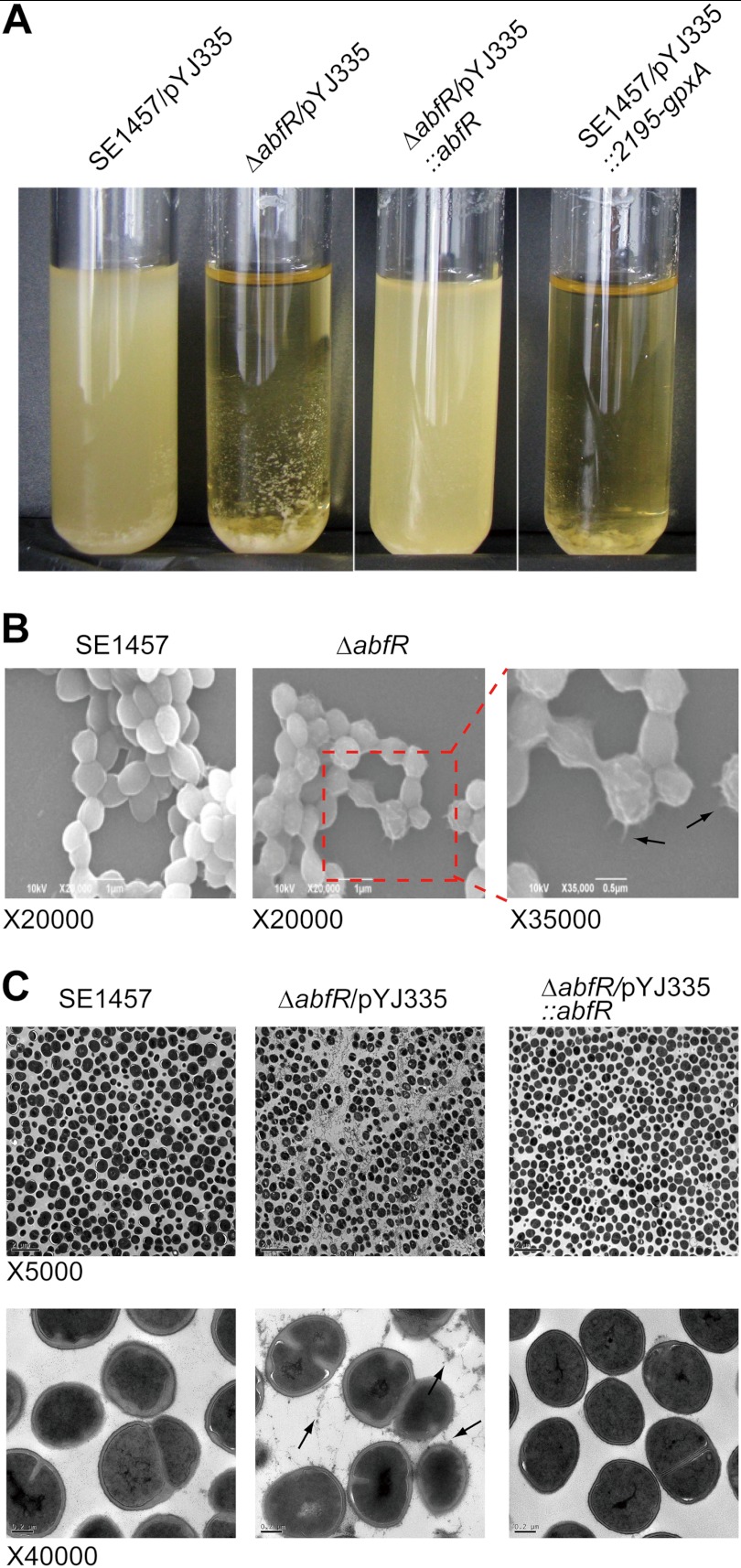
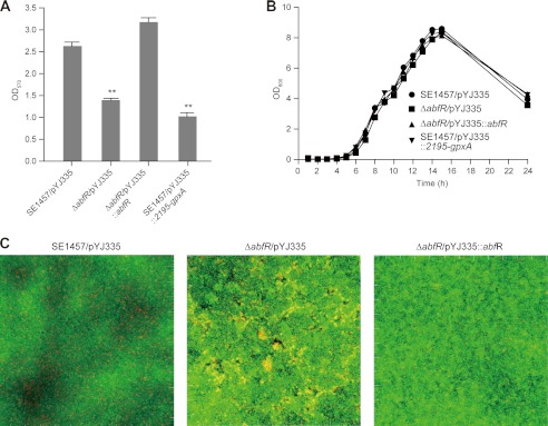
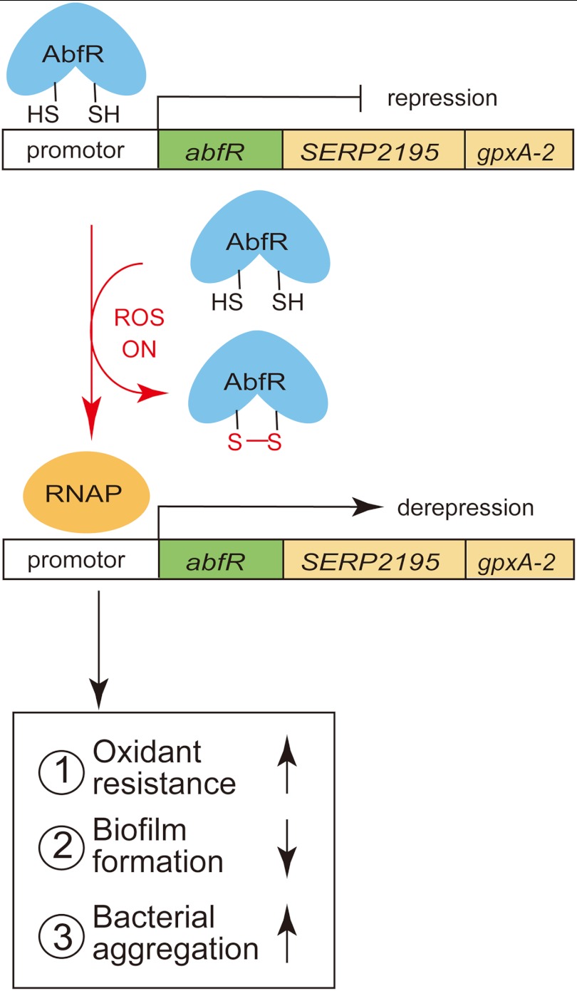
Staphylococcus epidermidis is a notorious human pathogen that is the major cause of infections related to implanted medical devices. Although redox regulation involving reactive oxygen species is now recognized as a critical component of bacterial signaling and regulation, the mechanism by which S. epidermidis senses and responds to oxidative stress remains largely unknown. Here, we report a new oxidation-sensing regulator, AbfR (aggregation and biofilm formation regulator) in S. epidermidis. An environment of oxidative stress mediated by H(2)O(2) or cumene hydroperoxide markedly up-regulates the expression of abfR gene. Similar to Pseudomonas aeruginosa OspR, AbfR is negatively autoregulated and dissociates from promoter DNA in the presence of oxidants. In vivo and in vitro analyses indicate that Cys-13 and Cys-116 are the key functional residues to form an intersubunit disulfide bond upon oxidation in AbfR. We further show that deletion of abfR leads to a significant induction in H(2)O(2) or cumene hydroperoxide resistance, enhanced bacterial aggregation, and reduced biofilm formation. These effects are mediated by derepression of SERP2195 and gpxA-2 that lie immediately downstream of the abfR gene in the same operon. Thus, oxidative stress likely acts as a signal to modulate S. epidermidis key virulence properties through AbfR.


Figures

FIGURE 1.
FIGURE 1.
Deduced structure and genomic environment of abfR in S. epidermidis. A, sequence alignment of SeAbfR, PaOspR, and SaMgrA was performed with Clustal Omega (67). The identical residues are highlighted in bold italic type. Asterisks indicate the cysteine residues. B, genetic context of the abfR locus is shown. The ORF number is indicated according to the S. epidermidis RP62A strain annotation. C, shown is co-transcription of abfR and gpxA-2. cDNA indicates co-transcriptional analyses of abfR and gpxA-2. RNA represents negative controls without reverse transcriptase. Genome represents positive controls using chromosomal DNA from S. epidermidis RP62A strain.
FIGURE 2.
FIGURE 2.
Gene expressions and phenotypes of S. epidermidis strains under oxidative stresses. Total RNA was extracted from S. epidermidis 1457 cultures (A600 = 0.8) treated with H2O2 or CHP for 30 min. A, qRT-PCR analyses of the transcripts of abfR in the presence of H2O2 and CHP, respectively. B, qRT-PCR analyses of the transcripts of SERP2195 and gpxA-2 under CHP stress. C, sensitivities of wild-type S. epidermidis 1457 strain harboring plasmid pYJ335 (SE1457/pYJ335), ΔabfR/pYJ335 strain, complemented strains (ΔabfR/pYJ335::abfR), and the SERP2195/gpxA-2 constitutive expression strain (SE1457/pYJ335::2195-gpxA) to oxidants. Exponential phase cells grown in BM medium were serially diluted (1:10 dilutions), and 10 μl of each dilution was spotted onto TSA agar containing either 8 mm H2O2 or 7 mm CHP. After incubation at 37 °C for ∼24 h, growth of the bacteria was observed. All of the experiments were performed in triplicate.
FIGURE 3.
FIGURE 3.
Quantitative PCR, gel shift assay, and DNase I footprint analysis. A, expression levels of SERP2195 and gpxA-2 in SE1457/pYJ335, ΔabfR/pYJ335, and ΔabfR/pYJ335::abfR were estimated by qRT-PCR. The experiment was replicated four times, and similar results were obtained. B, EMSA shows AbfR selectively bound to a 50-bp fragment DNA1, which included the putative −10 and −35 promoter regions. The sequence of DNAs 1–4 are listed in supplemental Table S2. C, AbfR binding site was determined by DNase I footprint assay. Electropherograms show the protection pattern of the abfR promoter in the absence or presence of 0.3 mm AbfR. The AbfR-protected region is indicated in the lower panel. D, the abfR promoter sequence (−31 to −8 from putative start codon GTG) is shown. The putative −35 and −10 promoter regions are underlined. Arrows show the palindromic AbfR-binding sequence that is highlighted in bold type within a box.
FIGURE 4.
FIGURE 4.
Role of cysteine residues in the regulatory function of AbfR. A, EMSA shows the binding between DNA1 and the reduced and oxidized AbfR proteins. CHP oxidation led to AbfR dissociation from the promoter DNA1. B, switching of EMSA binding between AbfR and DNA1 under the oxidized and reduced conditions. EMSA assays were performed on 50 nm DNA1 and 0.5 μm AbfR and 100 μm CHP or 1 mm DTT as indicated. A plus sign indicates the presence of a feature, and a minus sign indicates the absence of a feature. C, the binding of AbfR, AbfRC13S, and AbfRC116S to DNA1 under CHP exposure is shown. EMSA was performed on 50 nm DNA1, 0.5 μm protein, and CHP at varying concentration. D, DTNB analyses of the wild-type and mutant AbfR proteins treated or untreated with CHP are shown. E, qRT-PCR analyses of the transcripts of SERP2195 and gpxA-2 in a strain of ΔabfR/pYJ335::abfR, ΔabfR/pYJ335::abfRC13S, and ΔabfR/pYJ335:: abfRC116S, which were treated or untreated with CHP. F, plate assay showing sensitivities of S. epidermidis strains to CHP. Exponential phase cells grown in BM medium were serially diluted (1:10 dilutions), and 5 μl of each dilution was spotted onto TSA agar plate containing 9 mm CHP. The plates were incubated at 37 °C for ∼24 h.
FIGURE 5.
FIGURE 5.
Crystal structure of the reduced AbfR. A, crystal structure of AbfR in a dimeric dimmer is shown in cartoon. Two dimers existed in one asymmetric unit, in which one dimer is shown in magenta and pink and the other is shown in cyan and pale cyan. The distances between the intersubunit pairs Cys-13(′) and Cys-116(′) are shown, respectively. B, superimposition of the four monomers observed in A. The large conformational variation came from the dimerization motifs α1 and α6. C, one AbfR dimer is presented in cartoon. The secondary structural elements are shown (α, α-helices; β, β-sheets; wHTH motif, the helix-turn-helix wing region, β1-loop-β2). The distance between two α4 helices is ∼19 Å (defined as Cα of Asn-67). The distance between Cys-13 and Cys-116′ is 13 Å; these residues are favored for disulfide bond formation. D, secondary structure assignment of AbfR is shown. The residue numbers are shown on top of the sequence of AbfR, with the two Cys residues marked below with an asterisk. The secondary structural elements of the AbfR crystal structure (Protein Data Bank code 4HBL) are shown as red tubes for α-helices and blue arrows for β-sheets, respectively.
FIGURE 6.
FIGURE 6.
Characterization of disulfide bond formation in the oxidized AbfR. A–C, ultra performance liquid chromatography/electrospray ionization quadrupole-TOF mass spectrometric analysis extracted ion chromatograms of reduced AbfR (A), the oxidized AbfR (B), and the oxidized AbfR (C) treated with DTT after trypsin digestion. P1 indicates LANQLCFSAYNVSR, P2 indicates QLIITLTDNGQQQQEAVFEAISSCLPQEFDTTEYDETK, and P1 + P2 indicates cross-linked peptide between P1 and P2 through a disulfide bond. Nonreducing SDS-PAGE analyses of the reduced and oxidized AbfR samples are shown. D, electrospray ionization quadrupole-TOF mass spectrometric analysis mass spectrum of an unfractionated tryptic digestion. The quadruply charged (m/z 1478–1484) and triply charged (m/z 1970–1976) peaks, which are corresponding to the target disulfide-containing peptide (Theo Mr, theoretical mass: 5,915.7908 Da) are shown in a and b, respectively. Exp Mr, experimental mass. E, spectrum of the MSE fragmentation of the disulfide-containing peptide is shown. Y, y, B, b are types of ions. F, graphical fragment map that correlates the fragmentation ions to the peptide sequence. The disulfide-linked cysteines are shown in red and circled.
FIGURE 7.
FIGURE 7.
Phenotypes of S. epidermidis strains. A, bacterial aggregation of SE1457/pYJ335, ΔabfR/pYJ335, ΔabfR/pYJ335::abfR, and SE1457/pYJ335::2195-gpxA. The strains were grown in TSB at 37 °C for 12 h. ΔabfR/pYJ335 and SE1457/pYJ335::2195-gpxA cultures formed macroscopic aggregates that rapidly settled at the bottom of the test tubes. B, SEM images of SE1457 and ΔabfR are shown. It is presented at magnifications of ×20,000 and ×35,000, respectively. Arrows show the extracellular polymeric substances. C, transmission EM images of SE1457 and variants. Planktonic cells of SE1457, ΔabfR, and ΔabfR/pYJ335::abfR cultured for 16 h were examined by transmission EM. The images are displayed at magnification of ×5000 (top panels) and ×40,000 (bottom panels). Arrows show tuft-like surface material and extracellular polymeric substances between adjacent cells.
FIGURE 8.
FIGURE 8.
Effect of abfR deletion on biofilm formation. A, biofilm formation of SE1457/pYJ335, ΔabfR/pYJ335, ΔabfR/pYJ335::abfR, and SE1457/pYJ335::2195-gpxA are presented. The experiments were replicated four times, and similar results were obtained. The data are the means of 12 microtiter plate wells. Student's t test reveals significance. **, p value < 0.01. B, shown is the growth curve of SE1457/pYJ335, ΔabfR/pYJ335, ΔabfR/pYJ335::abfR, and SE1457/pYJ335::2195-gpxA. C, confocal laser scanning microscopy shows biofilm of S. epidermidis. Strains of SE1457, ΔabfR, and ΔabfR/pYJ335::abfR were incubated in glass-bottomed cell culture dishes. The results depict a stack of images taken at ∼0.3-μm-deep increments, and one of the three experiments is presented.
FIGURE 9.
FIGURE 9.
A proposed model of AbfR-based sensing and regulation of oxidative stress in S. epidermidis. Upon oxidative stress, AbfR senses reactive oxygen species (ROS) signals through the formation of the intersubunit disulfide bond and then dissociates from promoter DNA, which leads to derepression of the abfR-SERP2195-gpxA-2 operon. Consequently, the derepression of the SERP2195-gpxA-2 results in the reduced susceptibility to oxidant stress, decreased biofilm formation, and enhanced bacterial aggregation in S. epidermidis. RNAP, RNA polymerase.

Similar articles

Cited by

References

    1. Uçkay I., Pittet D., Vaudaux P., Sax H., Lew D., Waldvogel F. (2009) Foreign body infections due to Staphylococcus epidermidis. Ann. Med. 41, 109–119 - PubMed
    1. Götz F. (2002) Staphylococcus and biofilms. Mol. Microbiol. 43, 1367–1378 - PubMed
    1. Otto M. (2013) Staphylococcal infections. Mechanisms of biofilm maturation and detachment as critical determinants of pathogenicity. Annu. Rev. Med. 64, 1–14 - PubMed
    1. Imlay J. A. (2008) Cellular defenses against superoxide and hydrogen peroxide. Annu. Rev. Biochem. 77, 755–776 - PMC - PubMed
    1. Schlag S., Nerz C., Birkenstock T. A., Altenberend F., Götz F. (2007) Inhibition of staphylococcal biofilm formation by nitrite. J. Bacteriol. 189, 7911–7919 - PMC - PubMed

Publication types

MeSH terms

Associated data

LinkOut - more resources